Date: 26 November 2013
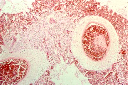
Pulmonary aspergillosis (K&E) (parrot C). Tissue from an individually housed and recently purchased, 6 month old African grey parrot found dead in the cage. Necropsy examination revealed focal necrosis of the left lung. This section stained by haematoxylin and eosin reveals septate fungal hyphae within the lung parenchyma. Similar hyphae were located in the walls and lumen of parabronchi, and within the walls of pulmonary blood vessels.
Copyright:
© Dr. Michael Day, University of Bristol
Notes: n/a
Images library
-
Title
Legend
-
Further details
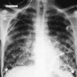
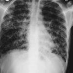
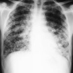
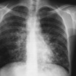
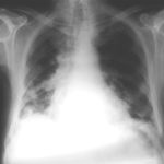
Image 1. The chest x-ray shows extensive bilateral nodular disease, most consistent with a fungal infection, or possibly tuberculosis. He was treated with a bucket face mask with 80% oxygen and voriconazole.
 ,
,  ,
,  ,
,  ,
,  ,
, 
-
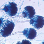
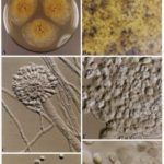
A Colonies on MEA +20 % sucrose after 2 weeks; B ascomata, x 40; C conidiophore of Aspergillus glaucus x 920;D conidiophore of Aspergillus glaucus x920 E. portion of ascoma with asci x 920. F ascospores x2330.
-
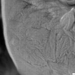
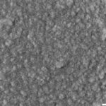
Scanning electron micrographs of A. fumigatus conidia of transformants rodB-02 (b). Size bar, 100 nm.

-
Scanning electron micrographs of A. fumigatus conidia of the wild-type G10 strain (a). Size bar, 100 nm.
-
Scanning electron micrographs of A. fumigatus conidia of rodA rodB-26 (d).Size bar, 100 nm.
-
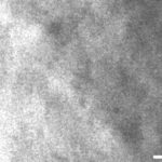
Scanning electron micrograph of an A.fumigatus conidium of rodA-47 (c), showing the hydrophobic rodlets covering the surface. Size bar, 100 nm.

 ,
, 
