Date: 26 November 2013
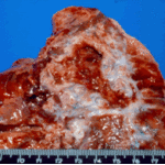
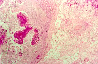
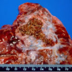
Pulmonary aspergillosis (cow 2). Section of lung from a 2 year old cow with weight loss and anorexia since calving. On necropsy examination, multiple firm masses were identified throughout the lungs. These were cavitating in nature, with a necrotic centre and peripheral fibrosis. Both this section and the following one are taken from the edge of such a lesion and demonstrate the pyogranulomatous inflammatory response.
Copyright:
© Dr. Michael Day, University of Bristol
Notes: n/a
Images library
-
Title
Legend
-
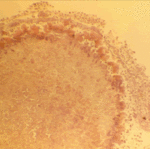
The periphery of the fungus ball is deeply eosinophilic because of the deposition of Splendore-Hoeppli material.
-
Single fungal ball, moving. Radiographic appearance of a fungus ball, showing movement as the patient’s position changes.
-
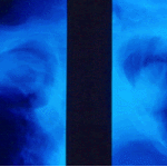
Oxalate crystals in the cavity wall surrounding an Aspergillus niger fungus ball (H&E, dark field, x 25).
-
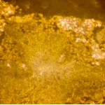
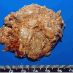
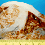
Aspergilloma patient. Gross pathology appearance of a fungus ball.
-
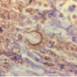
Conidiophores of Aspergillus fumigatus in the mass of the fungal ball surrounded by mycelia (H&E, x 400).
-
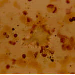
Aspergillus niger fungal ball. Calcium oxalate crystals in Aspergillus niger fungal ball. Also shown are darkly pigmented, rough-walled conidia associated with Aspergillus niger infection.
-
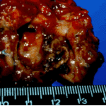
Aspergillus niger fungus ball within an old tuberculous cavern. This patient had diabetes, a disease commonly associated with A. niger infection.

 ,
, 
 ,
, 