Date: 26 November 2013
Further details
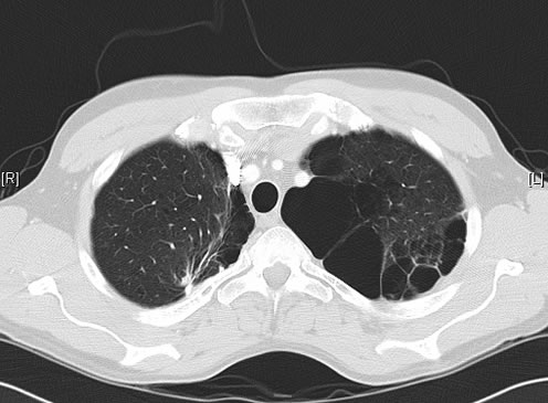
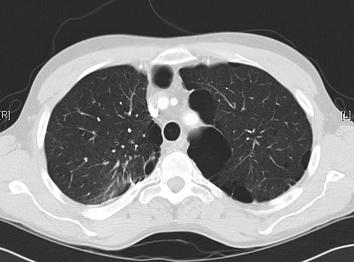
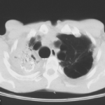
June 2009 – CT scan of the chest (two images D & E). These scans show residual large bullae, particularly notable in the apex of the left lung on both cuts. Small residual bullae remain in the right apex following a bullectomy which greatly reduced the size and number of these lesions.
Pt RK Lung bullae caused by cannabis smoking complicated by pneumothorax and chronic cavitary pulmonary aspergillosis.
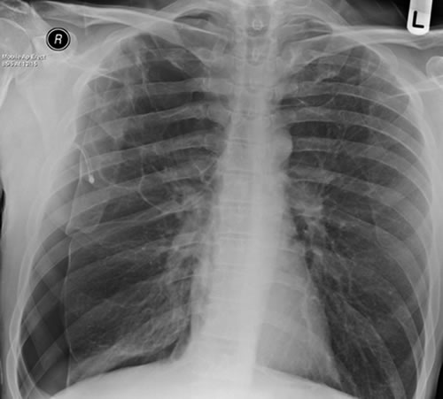
February 2009 – Chest x-ray showing bilateral apical bullae most marked on the right in association with a right pneumothorax. A chest drain is in-situ.
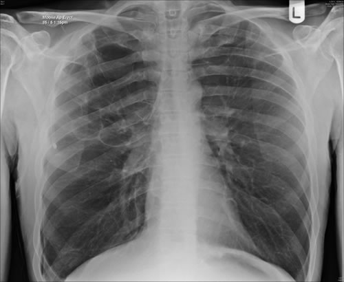
Late February 2009 – persistent apical bullae right upper zone with resolving pneumothorax. Chest drain still in-situ. Increasing right upper lobe shadowing possibly representing infection or haemorrhage.
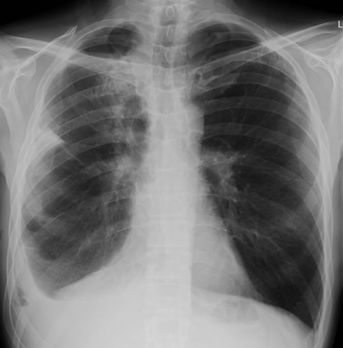
April 2009 – Post-operative chest x-ray showing post apical bullectomy on the right, resolved pneumothorax but the interval development of fluid in the right costo-phrenic angle with air fluid levels consistent with recent surgery. Great reduction in apical bullae in the right apex but increasing consolidation proximally in the right upper zone and some fluid in the horizontal fissure. These findings are following an apical bullectomy and pleurodesis of his prior significant pneumothorax.
June 2009 – CT scan of the chest (two images – see above). These scans show residual large bullae, particularly notable in the apex of the left lung on both cuts. Small residual bullae remain in the right apex following a bullectomy which greatly reduced the size and number of these lesions.
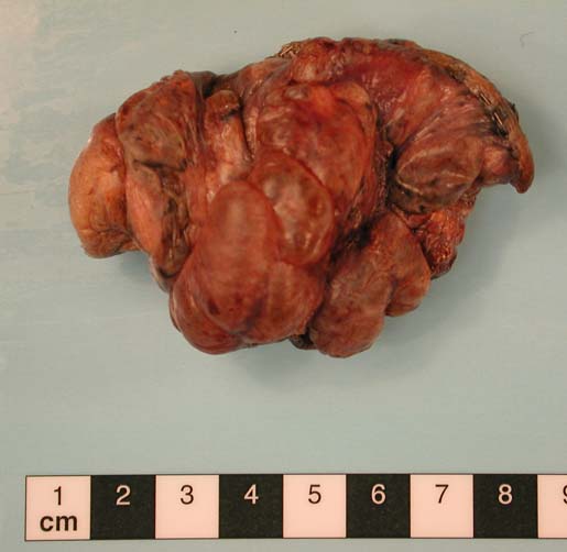
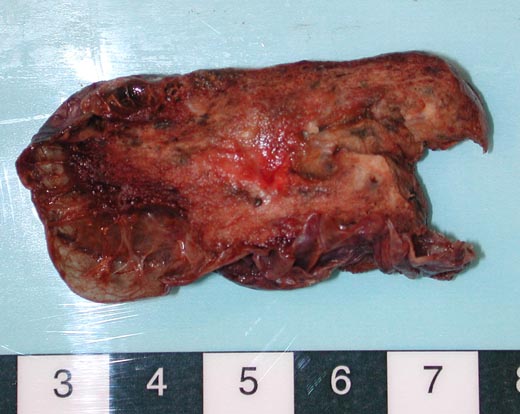
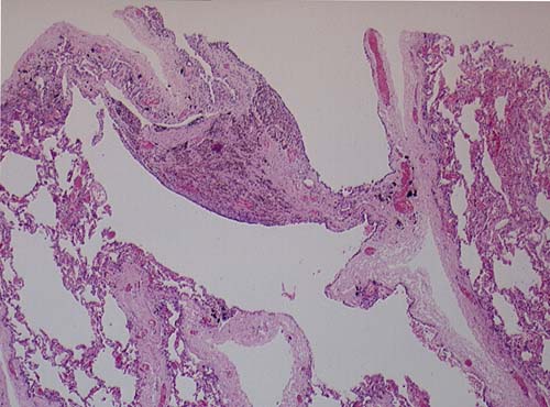
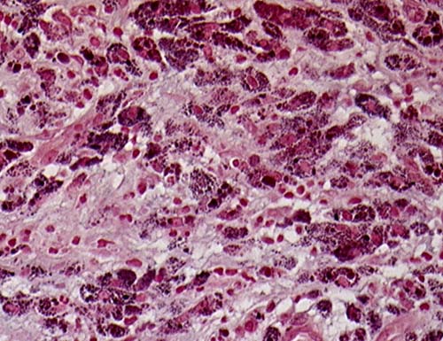
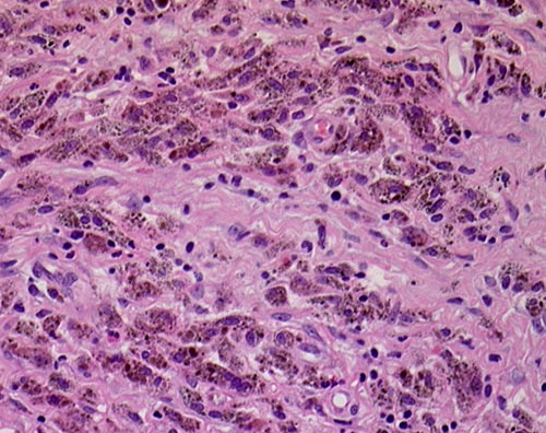
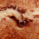
This patient underwent a bullectomy to remove part of the lung after developing lung bullae associated with cannabis smoking. The transverse section (G) of the specimen clearly shows the bullae along the left side and lower edge of the section. I and J demonstrate the dark pigment seen in accumulated macrophages. Tobacco smoking alone also causes a dark pigment, but evidence suggests that in marijuana smokers this pigment accumulates much faster and even with reduced exposure. The manner in which marijuana is smoked ie. without a filter and extended holding of the breath may contribute to this observation.
Copyright: n/a
Notes: n/a
Images library
-
Title
Legend
-
Diagnosis: Aspergilloma with invasive aspergillosis evidence of invasion found in the lumbar spine and brain, in addition to heart.
Fungal endocarditis with NO evidence of bacterial endocarditismia.Additional image details:
A. Normal chest X ray:
This (normal) chest X-ray was taken about 6 weeks before endocarditis was diagnosed, and 3 months before death due to disseminated aspergillosis. No CT scan was done (a chest radiograph has a 10% false negative rate in leukaemic patients, compared with CT).B. Aspergillus niger Fungal ball:
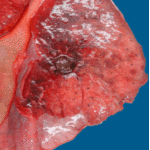
Gross section of lung at autopsy showing a discrete, well-demarcated dark/black mass surrounded by a fibrotic capsule. There was no evidence of local invasion, or infarction. The patient had had acute myeloid leukaemia (M1) and responded poorly to chemotherapy. He developed A.niger endocarditis and disseminated disease to the kidneys, lumbar disc and heart, probably arisiong from this lesion. It is unclear whether this lung lesion was a partially cured ‘mycotic lung sequestrum’ following antifungal therapy, or originated as an aspergilloma. The confirmation of genus and species was obtained by PCR on blood and vegetations.C. Endocarditis:
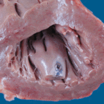
Macroscopic view of the heart at autopsy, showing an infracted lesion on the papillary muscle of the mitral valve in the left ventricle. In addition the patient had large vegetations, which are not shown here. The confirmation of genus and species was obtained by PCR on blood and vegetations; the pericarditis was a manifestation of disseminated aspergillosis.D. Pericarditis due to Aspergillus niger:
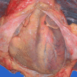
Macroscopic view of the pericardium at autopsy, showing gross chronic haemorrhagic pericarditis. The confirmation of genus and species was obtained by PCR on blood and vegetations; the pericarditis was a manifestation of disseminated aspergillosis.E. Lumbar discitis:
Macroscopic lesion of a lumbar intervertebral vertebral at autopsy, showing haemorrhagic necrosis, caused by hyphal invasion and infarction. The confirmation of genus and species was obtained by PCR on blood and vegetations; the discitis was a manifestation of disseminated aspergillosis.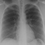 ,
,  ,
,  ,
,  ,
, 
-
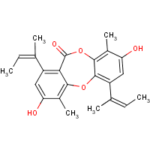
Secondary metabolites, structure diagram: Trivial name – Folipastatin
