Date: 26 November 2013
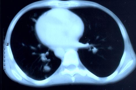
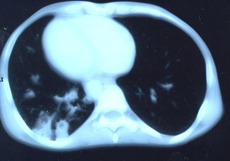
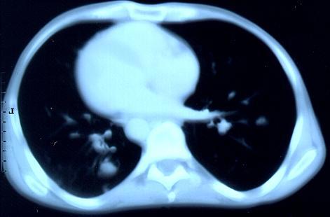
Transverse sections through the thorax of a patient with AIDS, hepatitis C and a left tempero-parietal cerebral lymphoma. His CD4 cell count was 45 x 106 / l. The lymphoma was proven by biopsy after a poor response to anti-toxoplasma therapy. He was given dexamethasone to cover the surgery and then developed diabetes mellitus. He did not receive chemotherapy for his lymphoma but did have 2 cerebral radiotherapy treatments (1.8 Gy each). Three weeks after the biopsy he developed dyspnoea and fever. Shortly after this he developed a right-sided hemiparesis, became comatose and died 2 days later.Autopsy showed a cerebral lymphoma and pulmonary and renal aspergillosis. Aspergillus nidulans was recovered from cultures of lungs and kidney.
Copyright:
Images submitted by Dr. Cornelia Lass-Floerl, University of Innsbruck – Institute of Hygiene; the case team includes: Dr. Mario Sarcletti, Dr. Alfons Stöger and Prof. Hans Maier all at the University of Innsbruck.
Notes: n/a
Images library
-
Title
Legend
-
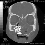
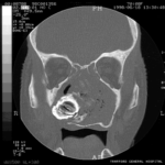
Patient MB X rays and CT scans. Chronic calcified maxillary sinusitis, patient had a palate defect.A. fumigatus cultured.
Images A&B Plain X rays antero-posterior and lateral, pre-operatively of Pt MB aged 76 who presented with unilateral nasal stuffiness and difficulty getting dentures fitted. She had hda these symptoms for many years. A large irregular calcified mass can be seen replacing the right maxillary sinus.
Images C D & E Coronal CT scan images of Pt MB showing a completely obstructed nasal cavity bilaterally and loss of internal nasal architecture. On the right side is large lamellar calcified lesion embedded in the extensive inflammatory material. Loss of bony margins is seen in numerous locations. This material was all removed surgically and showed mostly necrotic debris with Charcot-Leyden crystals and a few eosinophils and degenerate fungal hyphae. Aspergillus fumigatus was cultured from the material, especially infero-laterally on the right.
Image F Photograph through the mouth post-operatively showing the palate and a large defect in its right side. Through the defect can be seen the interior of the right maxillary sinus and nasal cavity with the inferior turbinate just visible.
 ,
,  ,
,  ,
,  ,
,  ,
, 

