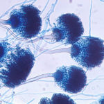
Date: 26 November 2013
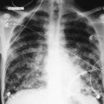
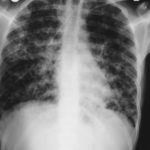
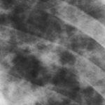
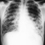
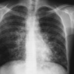
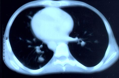
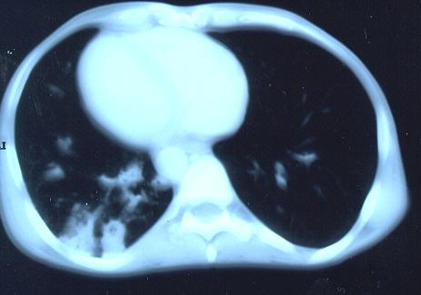
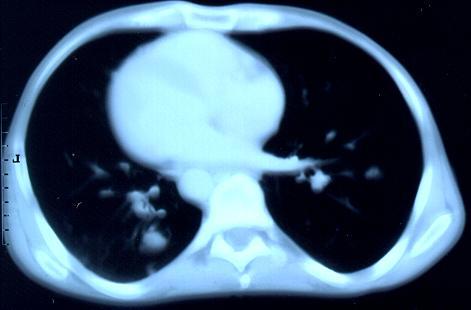
Transverse sections through the thorax of a patient with AIDS, hepatitis C and a left tempero-parietal cerebral lymphoma. His CD4 cell count was 45 x 106 / l. The lymphoma was proven by biopsy after a poor response to anti-toxoplasma therapy. He was given dexamethasone to cover the surgery and then developed diabetes mellitus. He did not receive chemotherapy for his lymphoma but did have 2 cerebral radiotherapy treatments (1.8 Gy each). Three weeks after the biopsy he developed dyspnoea and fever. Shortly after this he developed a right-sided hemiparesis, became comatose and died 2 days later.Autopsy showed a cerebral lymphoma and pulmonary and renal aspergillosis. Aspergillus nidulans was recovered from cultures of lungs and kidney.
Copyright:
Images submitted by Dr. Cornelia Lass-Floerl, University of Innsbruck – Institute of Hygiene; the case team includes: Dr. Mario Sarcletti, Dr. Alfons Stöger and Prof. Hans Maier all at the University of Innsbruck.
Notes: n/a
Images library
-
Title
Legend
-
Further details
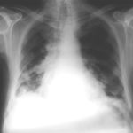
Image 1. The chest x-ray shows extensive bilateral nodular disease, most consistent with a fungal infection, or possibly tuberculosis. He was treated with a bucket face mask with 80% oxygen and voriconazole.
 ,
,  ,
,  ,
,  ,
,  ,
, 
-
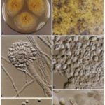
A Colonies on MEA +20 % sucrose after 2 weeks; B ascomata, x 40; C conidiophore of Aspergillus glaucus x 920;D conidiophore of Aspergillus glaucus x920 E. portion of ascoma with asci x 920. F ascospores x2330.
-
Scanning electron micrographs of A. fumigatus conidia of transformants rodB-02 (b). Size bar, 100 nm.

-
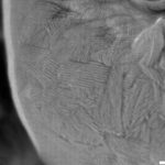
Scanning electron micrographs of A. fumigatus conidia of the wild-type G10 strain (a). Size bar, 100 nm.
-
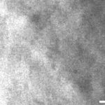
Scanning electron micrographs of A. fumigatus conidia of rodA rodB-26 (d).Size bar, 100 nm.
-
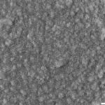
Scanning electron micrograph of an A.fumigatus conidium of rodA-47 (c), showing the hydrophobic rodlets covering the surface. Size bar, 100 nm.


 ,
, 


