Date: 26 November 2013
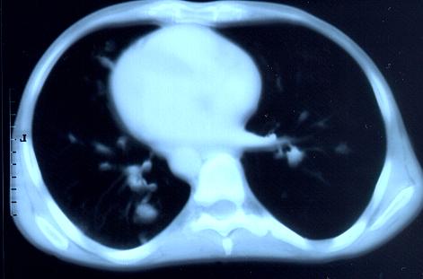
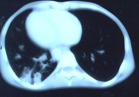
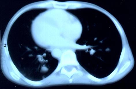
Transverse sections through the thorax of a patient with AIDS, hepatitis C and a left tempero-parietal cerebral lymphoma. His CD4 cell count was 45 x 106 / l. The lymphoma was proven by biopsy after a poor response to anti-toxoplasma therapy. He was given dexamethasone to cover the surgery and then developed diabetes mellitus. He did not receive chemotherapy for his lymphoma but did have 2 cerebral radiotherapy treatments (1.8 Gy each). Three weeks after the biopsy he developed dyspnoea and fever. Shortly after this he developed a right-sided hemiparesis, became comatose and died 2 days later.Autopsy showed a cerebral lymphoma and pulmonary and renal aspergillosis. Aspergillus nidulans was recovered from cultures of lungs and kidney.
Copyright:
Images submitted by Dr. Cornelia Lass-Floerl, University of Innsbruck – Institute of Hygiene; the case team includes: Dr. Mario Sarcletti, Dr. Alfons Stöger and Prof. Hans Maier all at the University of Innsbruck.
Notes: n/a
Images library
-
Title
Legend
-
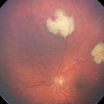
Bilateral A. fumigatus endophthalmitis in association with pulmonary and cerebral aspergillosis, complicating severe autoimmune disease treated with intense immunosuppression. Left eye.
-
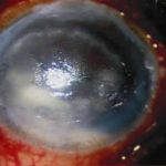
Aspergillus keratitis – a case of fungal keratitis following amnoiotic membrane transplantation (AMT) for bullous keratopathy. Slit-lamp photograph of left eye showing ring shaped stromal infiltrate.
-
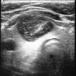
A 14 year old boy with a T cell lymphoma received induction chemotherapy with high dose dexamethasone. At no time during this therapy was he neutropenic. Three weeks into treatment his dexamethasone was reduced and stopped due to gastro-intestinal side effects. On recovery of his abdominal symptoms, he developed sudden onset of severe right sided pleuritic chest pain. Initially, he was thought to have a pulmonary embolism as a chest X-ray showed a solid wedge shaped area of consolidation. He then developed a cough and temperature and sputum grew Aspergillus fumigatus. The wedge shaped lesion developed cavitation. Despite AmBisome at 5mg/Kg, commenced within 4 days of onset of symptoms, the chest X-ray appearance got gradually worse over the following week. This led to substitution of Ambisome by caspofungin and voriconazole. He developed a thyroid cyst with haemorrhage that on aspiration grew A. fumigatus. His chest X-ray continued to worsen. He underwent a right lower lobe lobectomy, which confirmed the diagnosis of invasive pulmonary aspergillosis. Unfortunately the thyroid cyst continued to increase in size resulting in removal of the right lobe of the thyroid.
Chest CT scan carried out 3 weeks after lobectomy revealed new lesions within the right upper lobe of the lung. At this time the voriconazole therapy was stopped and posaconazole started. The area of the thyroid remains free of Aspergillus and the lung lesions appear to be improving 6 weeks after the start of posaconazole.
 ,
,  ,
, 
-
Aspergillus keratitis. Severe peripheral lesion due to aspergillus unlikely to respond well to treatment.

-
Aspergillus keratitis. Corneal scar at end of treatment of case in previous slide.Vision recovered to 6/9.

-
Aspergillus keratitis. Severe central aspergillus infection with a “cheesey†looking area of the lesion and hypopyon (fluid level of inflammatory cells in the anterior chamber)

-
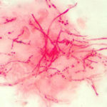
Corneal ulcer – gram stain. Corneal scrapings were taken from a 67 yr old farmer presenting with a corneal ulcer of the right eye. A piece of vegetable matter was embedded in the cornea and scrapings were done. Gram stain (500x magnification) showed numerous septate hyphae. Cultures grew a small amount of A fumigatus.
