Date: 21 January 2014
Further details
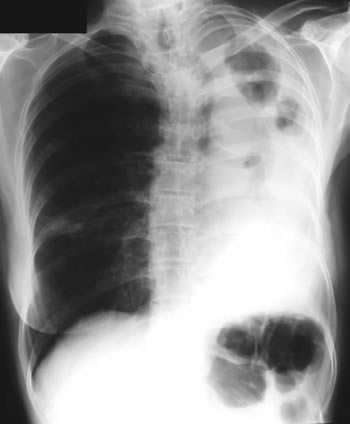
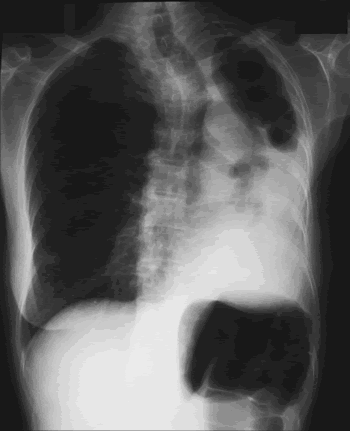
Image B. Additional cavities are apparent inferior to this large cavity and are in communication both with the bronchi and the additional cavities. Some of the apparent cavities are probably dilated bronchi. The left lower lung is completely opacified otherwise. The degree of pleural fibrosis surrounding the left apical cavity is reduced slightly over the interval of four months.
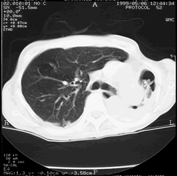
Image C. This shows an almost normal hyperexpanded right lung with a very substantially contracted left lung with one large airway visible and probably incontinuity with a slightly irregular cavity containing some debris, presumably fungal tissue. Other levels show very large left apical cavity with numerous subsections containing debris or fibrotic tissue and almost complete fibrosis of the lung below the level of the carina on the left, with some calcification within the fibrotic lung tissue.
Copyright: n/a
Notes: n/a
Images library
-
Title
Legend
-
Saggital section of the vertebral column of a dog with discospondylitis as part of disseminated aspergillosis due to A. terreus

-
Discospondylitis – Radiograph of thoracolumbar vertebrae of a 2 year old, female German shepherd dog with disseminated aspergillosis demonstrating discospondylitis of T13-L1.
-
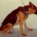
Wasting and paraplegia – A 2 year old, male German shepherd dog with disseminated aspergillosis due to A. terreus. The marked loss of condition of this dog occurred within two months of initial diagnosis.
-
A 2 year old, female German shephered dog with disseminated aspergillosis due to A. terreus. There is muscle wasting and paraplegia due to discospondylitis involving T13-L1.

-
Draining sinus tract on left forelimb of a 4 year old, female Dalmatian with disseminated aspergillosis. There was an underlying osteomyelitis of distal humerus from which A. terreus was cultured.

-
Aspergillus sinusitis in a dog. Long nosed dogs are at relatively high risk of Aspergillus sinusitis as shown in this example.

-
English Pointer with nasal aspergillosis. Gram stained cytological smear of material obtained from the frontal sinus of a 7 year old English Pointer with nasal aspergillosis. This infection was caused by Aspergillus fumigatus. Magnification x 200.

-
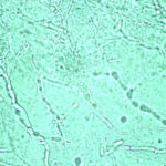
Domestic crossbred cat with disseminated aspergillosis. KOH preparation of material obtained from thoracotomy of a 3 year old domestic crossbred cat with invasive Aspergillus fumigatus infection. The cat had marked enlargement of the hilar lymph nodes that resulted in a partial tracheal obstruction. This preparation was made from portions of the hilar lymph node resected at thoracotomy. Magnification x 132.
-
Rottweiler treated with indwelling plastic tubes. Photograph of a Rottweiler crossbred dog treated with indwelling plastic tubes placed surgically into the nasal cavity and frontal sinuses.
-
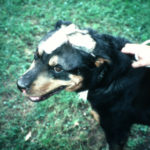
Nasal aspergillosis in an English Pointer. Photograph of an English Pointer with nasal aspergillosis
Nasal aspergillosis in an English Pointer. Photograph of an English Pointer with nasal aspergillosis


