Date: 21 January 2014
Further details
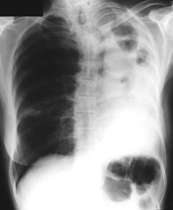
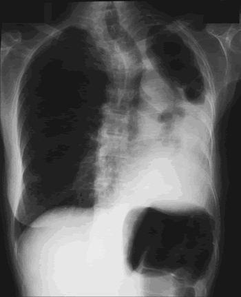
Image B. Additional cavities are apparent inferior to this large cavity and are in communication both with the bronchi and the additional cavities. Some of the apparent cavities are probably dilated bronchi. The left lower lung is completely opacified otherwise. The degree of pleural fibrosis surrounding the left apical cavity is reduced slightly over the interval of four months.
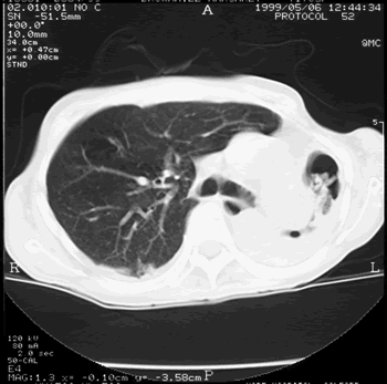
Image C. This shows an almost normal hyperexpanded right lung with a very substantially contracted left lung with one large airway visible and probably incontinuity with a slightly irregular cavity containing some debris, presumably fungal tissue. Other levels show very large left apical cavity with numerous subsections containing debris or fibrotic tissue and almost complete fibrosis of the lung below the level of the carina on the left, with some calcification within the fibrotic lung tissue.
Copyright: n/a
Notes: n/a
Images library
-
Title
Legend
-
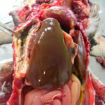
Aspergillosis in penguins. Lesions found in captive Magellanic penguins (Spheniscus magellanicus) with aspergillosis as determined by histology. Adherence areas of air sac to the celomic wall and white-yellowish nodule in the liver.

-
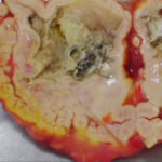
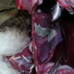
Aspergillosis in penguins. Lesions found in fatal cases of captive Magellanic penguins (Spheniscus magellanicus) with aspergillosis. Air sacs thickened with abundant plaque-like caseous and necrotic debris covering the wall with greyish-green fungal colonies on the internal surface.
-
Aspergillosis in penguins. Lesions found in fatal cases of captive Magellanic penguins (Spheniscus magellanicus) with aspergillosis. Air sacs thickened with abundant plaque-like caseous and necrotic debris covering the wall with greyish-green fungal colonies on the internal surface.
-
Aspergillosis in penguins. Lesions found in fatal cases of captive Magellanic penguins (Spheniscus magellanicus) with aspergillosis. Air sacs thickened with abundant plaque-like caseous and necrotic debris covering the wall with greyish-green fungal colonies on the internal surface.
-
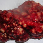
Aspergillosis in penguins. Lesions found in captive Magellanic penguins (Spheniscus magellanicus) with aspergillosis.Lung parenchyma with congestion, hemorrhagic and necrotic areas and with multiple white-yellowish granulomatous nodules, ranging from 0.1-1.0 cm in diameter

-
Aspergillosis in penguins. Lesions found in captive Magellanic penguins (Spheniscus magellanicus) with aspergillosis. Lung parenchyma with congestion, hemorrhagic and necrotic areas and with multiple white-yellowish granulomatous nodules, ranging from 0.1-1.0 cm in diameter
-
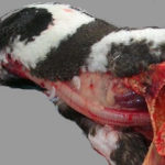
Aspergillosis in penguins. Lesions found in captive Magellanic penguins (Spheniscus magellanicus) with aspergillosis.
