Date: 26 November 2013
The patient was a 610 g twin male born by spontaneous normal vaginal delivery at 23 weeks and 4 days gestation. He was started on benzyl penicillin and gentamicin for sepsis. On day 3, he developed metabolic acidosis, hyponatremia, anemia, thrombocytopenia and jaundice and his antibiotics were changed to vancomycin, cefotaxime and fluconazole.
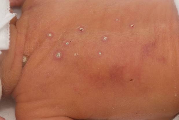
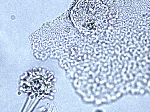
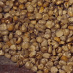
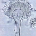
On day 10, multiple circular skin papules with white eschars were noted on his back (Figure A). A full septic screen was repeated including skin scraping and biopsy for urgent microscopy and culture. Microscopy of skin scrapes revealed fungal elements including hyphae and fruiting heads suggestive of Aspergillus spp (Figure B). Lipid amphotericin B was commenced and fluconazole was stopped. Skin scrapings on culture grew Aspergillus fumigatus. A diagnosis of primary cutaneous aspergillosis was made. The patient responded to oral posaconazole 6mg/kg/8 hourly. All lesions disappeared after 44 days and he continued with posaconazole until day 60.
Published case at Langan et al Pediatr Dermatol 2010 Jul-Aug 27 (4) 403-4
Copyright: n/a
Notes:
Images library
-
Title
Legend
-
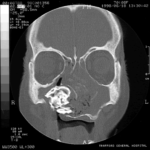
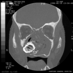
Patient MB X rays and CT scans. Chronic calcified maxillary sinusitis, patient had a palate defect.A. fumigatus cultured.
Images A&B Plain X rays antero-posterior and lateral, pre-operatively of Pt MB aged 76 who presented with unilateral nasal stuffiness and difficulty getting dentures fitted. She had hda these symptoms for many years. A large irregular calcified mass can be seen replacing the right maxillary sinus.
Images C D & E Coronal CT scan images of Pt MB showing a completely obstructed nasal cavity bilaterally and loss of internal nasal architecture. On the right side is large lamellar calcified lesion embedded in the extensive inflammatory material. Loss of bony margins is seen in numerous locations. This material was all removed surgically and showed mostly necrotic debris with Charcot-Leyden crystals and a few eosinophils and degenerate fungal hyphae. Aspergillus fumigatus was cultured from the material, especially infero-laterally on the right.
Image F Photograph through the mouth post-operatively showing the palate and a large defect in its right side. Through the defect can be seen the interior of the right maxillary sinus and nasal cavity with the inferior turbinate just visible.
 ,
,  ,
,  ,
,  ,
,  ,
, 

