Date: 26 November 2013
Born 75 years ago, Pt HK had 3 episodes of tuberculosis as a child and teenager, being treated with PAS and streptomycin. He suffered a ‘bad chest’ all his life and retired aged 54. Presenting with worsening and more frequent chest infections, he was referred with ‘bronchiectasis and Aspergillus sensitisation’. A diagnosis of chronic pulmonary aspergillosis was made in June 2009 on the basis of his chest radiograph and strongly positive Aspergillus precipitins (IgG antibodies) (titre 1/16). He also had Pseudomonas aeruginosa colonisation. His oxygen saturation was 87% and his pO2 6.8, pCO2 6.2 KPa.
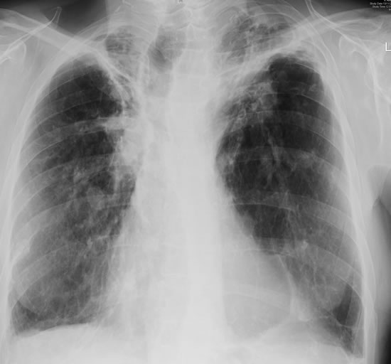
His chest radiograph (see above, November 2009) was reported as showing; “ The lung fields are over-inflated. Bilateral apical fibrotic change secondary to old TB. No cavity seen.” At clinic, bilateral apical cavities were seen, with some associated pleural thickening at the left apex, without any evidence of a fungal ball.
He started posaconazole 400mg twice daily with therapeutic levels at subsequent visits. Sputum cultures never grew Aspergillus. Over the following 9 months he had no chest infections requiring antibiotics, his breathlessness worsened gradually and he remained easily fatigued. His Aspergillus antibody titres fell. Overall he felt better, but was concerned about declining respiratory status.
Copyright:
Fungal Research Trust
Notes: n/a
Images library
-
Title
Legend
-
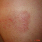
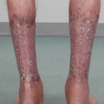
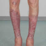
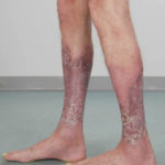
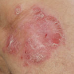
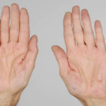
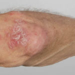
After 3 weeks of posaconazole given for chronic pulmonary aspergillosis, patient NC had a remarkable exacerbation of psoriasis. He had had psoriasis for years, with little trouble and almost no treatment. After taking posaconazole 400mg twice daily, he developed psoriatic plaques on his hands for the first time ever. The plaques on his lower legs became confluent. This occurred in association with worsening chest symptoms, notably increased coughing, more breathlessness and increasing oxygen requirement.
Posaconazole was stopped after 3 weeks, and 2 weeks later he was still very symptomatic with his chest. This responded to a 2 week course of corticosteroids, and his psoriasis also improved.
 ,
,  ,
,  ,
,  ,
,  ,
, 
-
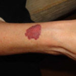
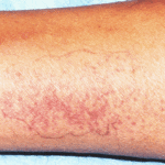
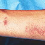
Patient PC: An example of localised caspofungin rash and phlebitis related to caspofungin infusion.
 ,
, 
-
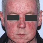
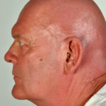
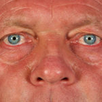
This 55 year old man with asthma, ABPA, severe bronchiectasis and lung fibrosis was treated with voriconazole, starting in June 2010. He had developed increasing dyspnoea on itraconazole for over 7 years, and his total IgE remained at 1100 KIU/L. He had marked photopsia (visual hallucinations) and facial erythema in the first 3 weeks of therapy. His trough voriconazole concentration was 1.17 mg/L. Over 3 months, he had minor improvement in his breathlessness but continued facial erythema, despite factor 50 sunblock. After 5 months of therapy his facial rash has altered to show acneiform lesions with localised crusting and background severe erythema. His face effectively crusted over, and he stopped therapy.
Over the next 3 weeks his facial appearance slowly improved .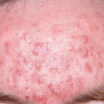 ,
,  ,
, 