Date: 26 November 2013
Born 75 years ago, Pt HK had 3 episodes of tuberculosis as a child and teenager, being treated with PAS and streptomycin. He suffered a ‘bad chest’ all his life and retired aged 54. Presenting with worsening and more frequent chest infections, he was referred with ‘bronchiectasis and Aspergillus sensitisation’. A diagnosis of chronic pulmonary aspergillosis was made in June 2009 on the basis of his chest radiograph and strongly positive Aspergillus precipitins (IgG antibodies) (titre 1/16). He also had Pseudomonas aeruginosa colonisation. His oxygen saturation was 87% and his pO2 6.8, pCO2 6.2 KPa.
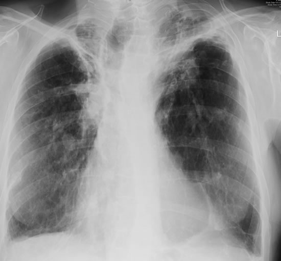
His chest radiograph (see above, November 2009) was reported as showing; “ The lung fields are over-inflated. Bilateral apical fibrotic change secondary to old TB. No cavity seen.” At clinic, bilateral apical cavities were seen, with some associated pleural thickening at the left apex, without any evidence of a fungal ball.
He started posaconazole 400mg twice daily with therapeutic levels at subsequent visits. Sputum cultures never grew Aspergillus. Over the following 9 months he had no chest infections requiring antibiotics, his breathlessness worsened gradually and he remained easily fatigued. His Aspergillus antibody titres fell. Overall he felt better, but was concerned about declining respiratory status.
Copyright:
Fungal Research Trust
Notes: n/a
Images library
-
Title
Legend
-
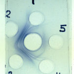
Aspergillus – Immunodiffusion Test showing stong immunoprecipitin lines against aspergillus.
-
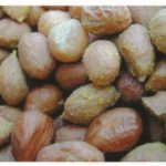
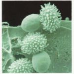
Talaromyces macrosporus- very short heat treatments of dormant ascospores at 85 C are able to activate these spores to germinate within 1 minute. One stage of germination is a very quick bursting of the thinly walled inner cell through the thick ornamented outer cell wall of the spore.
-
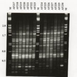
RAPD profiles of 14 A fumigatus isolates from Swedish and New Zealand saw mills, generated with primer R108. Mwt markers (lambda with Pst1) indicated by M.
-
These 3 images show non-union of the sternum post-aortic valve replacement as a result of local Aspergillus fumigatus infection. Underlying the sternum is some soft tissue which is presumptively also infection and in the most inferior image, the pericardium is also invoved.
The patient also is diabetic, with rhematoid arthritis and ulcerative colitis and occasionally receives a course of corticosteroids. The wound discharged pus which grew A. fumigatus. It was managed conservatively with itraconazole initally, with failure of therapy over 3 months.
 ,
,  ,
, 
-
CHEF gel of A. fumigatus chromosomes. The electrophoretic karyotype of two isolates of A. fumigatus is shown. The chromosomal bands have been resolved using Bio-Rad CHEF DRII equipment. The sizes of the bands vary between the two isolates and at least five bands have been resolved using the following conditions: chromosomal grade agarose (Bio-Rad) at 1.2 % in 1 x TAE buffer was used; the gel was run with a field strength of 1.8 V/cm for 48 h with a switch time of 2200 s and for 71 h with a switching ramp

-
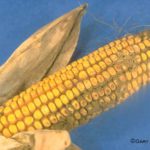
Aspergillus ear rot and storage mould – Aspergillus flavus and Aspergillus parasiticus can produce aflatoxins are generally known as storage fungi, but they can also cause ear rots in the field. These species are observed as a gray-green, powdery molds and they can be detected in corn because they produce compounds that are fluorescent under black light.
-
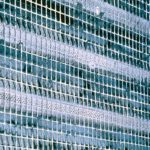
Environmental and sick building images. Dirty ventilation intake ducts
-
A.fumigatus conidium. Scanning electron micrograph of an A.fumigatus conidium showing the fascicles of hydrophobic rodlets covering the surface


