Date: 26 November 2013
Born 75 years ago, Pt HK had 3 episodes of tuberculosis as a child and teenager, being treated with PAS and streptomycin. He suffered a ‘bad chest’ all his life and retired aged 54. Presenting with worsening and more frequent chest infections, he was referred with ‘bronchiectasis and Aspergillus sensitisation’. A diagnosis of chronic pulmonary aspergillosis was made in June 2009 on the basis of his chest radiograph and strongly positive Aspergillus precipitins (IgG antibodies) (titre 1/16). He also had Pseudomonas aeruginosa colonisation. His oxygen saturation was 87% and his pO2 6.8, pCO2 6.2 KPa.
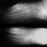
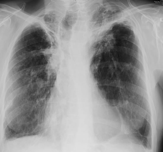
His chest radiograph (see above, November 2009) was reported as showing; “ The lung fields are over-inflated. Bilateral apical fibrotic change secondary to old TB. No cavity seen.” At clinic, bilateral apical cavities were seen, with some associated pleural thickening at the left apex, without any evidence of a fungal ball.
He started posaconazole 400mg twice daily with therapeutic levels at subsequent visits. Sputum cultures never grew Aspergillus. Over the following 9 months he had no chest infections requiring antibiotics, his breathlessness worsened gradually and he remained easily fatigued. His Aspergillus antibody titres fell. Overall he felt better, but was concerned about declining respiratory status.
Copyright:
Fungal Research Trust
Notes: n/a
Images library
-
Title
Legend
-
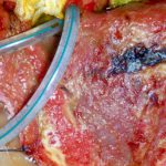
This patient, had had a laparostomy for recurrent intra-abdominal sepsis following on from crohns disease. She was transferred to another intensive care unit and her dressings changed daily. Shortly after, this dark patches appeared on her liver (as seen here A) and her colon. Superficial biopsies and culture showed A.fumigatus invading liver capsule. She responded to amphotericin B therapy.
B shows patient after treatment.
 ,
, 
-
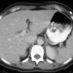
Hepatic aspergillosis, pt KO. Repeat CT scan of the liver showing almost complete resolution of lesions on itraconazole therapy.
-
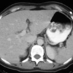
Image A. The CT scan of her abdomen had the appearances shown here. She also has small pulmonary nodules. Bioposy of the liver revealed hyphae consistent with Aspergillus.
Image B. She responded well to oral itraconazole therapy.
 ,
, 
-
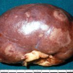
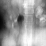
This image shows the pelvis of the left kidney filled with fungal balls. Eventually, after failing amphotericin B therapy, she required a nephrectomy. Her case is reported in Davies SP, Webb WJS, Patou G, Murray WK, Denning DW. Renal aspergilloma – a case illustrating the problems of medical therapy. Nephrol Dial Transplant 1987; 2: 568-572.
-
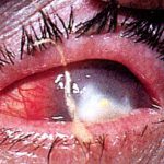
Aspergillus keratitis. Good example of Aspergillus keratitis caused by A.glaucus. Usually A.fumitagus and A.flavus are the causes.

 ,
, 