Date: 23 January 2014
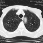
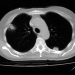
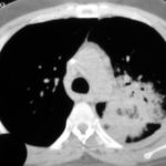
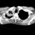
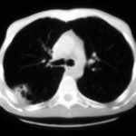
A 43 year old with smoking related emphysema was admitted to hospital with two separate episodes of haemoptysis. He had been in good health up to 1989, when he was diagnosed as having bilateral pulmonary tuberculosis. At that time a CT scan revealed a cavity in the left upper lobe (20.8cm2) with adjacent confluent infiltrates and pleural thickening. On bronchoscopic examination no abnormalities were noted and endobronchial biopsies did not reveal hyphae.
Over the next 4 years his condition deteriorated and a CT scan showed the left upper lobe cavity had increased to 40cm2. Itraconazole 400mg daily was prescribed. There was some clinical improvement on itraconazole but patient eventually deteriorated with breathlessness and with significant weight loss.
Copyright: n/a
Notes:
Images library
-
Title
Legend
-
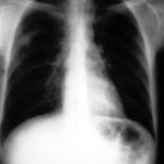
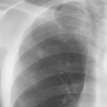
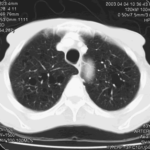
Nodules and areas of atelectasis are seen at both bases. He later died.

-
Further details
It is clearly a relatively small cavitary lesion, and the patient was almost asymptomatic. This response was a ‘stable’ response. The patient was included in the report Denning DW, Lee JY, Hostetler JS, Pappas P, Kauffman CA, Dewsnup DH, Galgiani JN, Graybill JR, Sugar AM, Catanzaro A, Gallis H, Perfect JR, Dockery B, Dismukes WE, Stevens DA, NIAID Mycoses Study Group multicenter trial of oral itraconazole therapy of invasive aspergillosis. Am J Med 1994; 97: 135-144.
 ,
, 
-
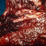
Well demarcated pulmonary infarction is well seen in this close-up of the lung at autopsy in a patient with histologically confirmed invasive aspergillosis. Angio invasion is characteristic of invasive aspergillosis, is associated with a worse prognosis, but is not always seen.
-
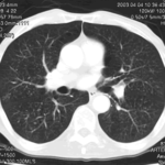
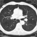
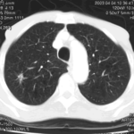
This 83 year old man presented with weight loss to a lung cancer clinic in mid 2003.
 ,
,  ,
,  ,
,  ,
,  ,
, 