Date: 26 November 2013
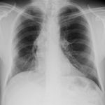
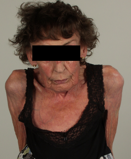
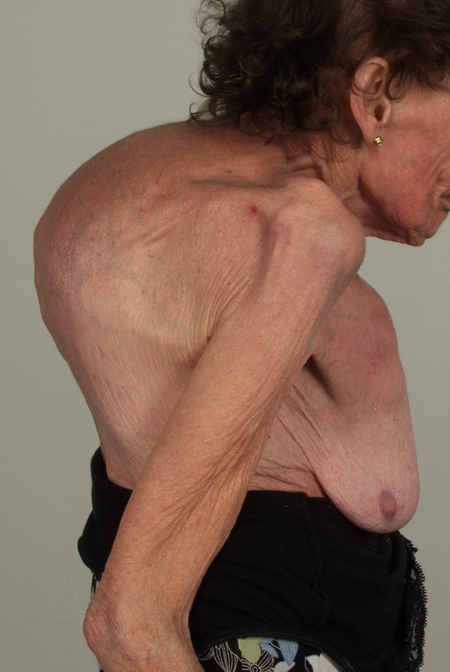
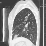
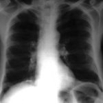
These pictures show remarkable curvature of the spine as a result of collapse of the vertebral bodies of the thoracic vertebrae. This is a gross example of steroid-induced osteoporosis. The dose was not large in the last 10 years, typically 5-10mg daily, but multiple high dose courses and slow tapering lead to this outcome.
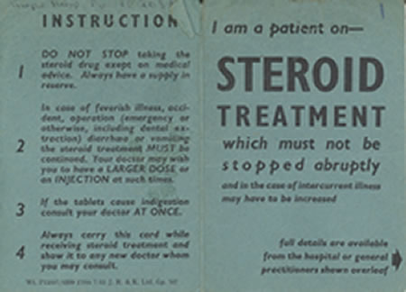
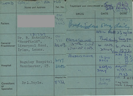
Her corticosteroid warning card is also demonstrated, as additional steroids are required for any significant illness or surgery, as her adrenal glands had completely atrophied.
Kindly supplied by Prof David Denning, South Manchester University Hospitals NHS Trust, Manchester UK
(© Fungal Research Trust)
Copyright:
Kindly supplied by Prof David Denning, South Manchester University Hospitals NHS Trust, Manchester UK
Notes:
Images library
-
Title
Legend
-
pt FT. Normal chest radiograph of patient with extensive pseudomembranous Aspergillus tracheobronchitis, 4 days before death.
-
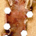
Pt FT. Autopsy appearance of the trachea, after the adherent pseudomembrane had been removed, revealing confluent ulceration superiorly with small green plaques of Aspergillus growth on the trachea inferiorly.
-
This view was obtained in a lung transplant recipient at bronchoscopy. Aspergillus fumigatus was grown from bronchial lavage but invasion was not demonstrated on bronchial biopsy. Symptoms improved with itraconazole therapy and abnormal appearances had resolved within 2 weeks.

-
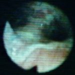
Bronchoscopic view of Aspergillus tracheobronchitis. Bronchial lavage revealed hyphae in microscopy and cultures grew A.fumigatus. This man had received a lung transplant a few weeks before. Invasion of mucosa, but not cartilage, was demonstrated histologically. He responded rapidly to oral itraconazole.
-
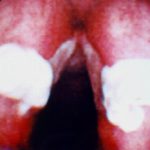
This view from indirect laryngoscopy illustrates bilateral lesions on the larynx that on biopsy were shown to be due to Aspergillus. This is a rare disease in non-immunocompromised patients.
-
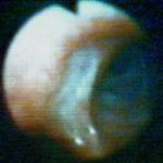
Bronchoscopic view of a deep bronchial ulcer in a lung transplant patient. Biopsies through the ulcer yielded cartilage with hyphae invading it. Fungal cultures of bronchial lavage grew Aspergillus fumigatus. He responded to oral itraconazole therapy.
-
Patient had life threatening pneumonia, cavity formation was later observed. He later presented with a fungal ball. The aspergilloma was removed by surgical resection of the right upper lobe.
 ,
,  ,
,  ,
,  ,
,  ,
,  ,
, 