Date: 26 November 2013
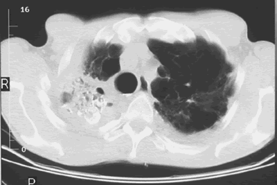
Image A. Scan shows large bore needle in one of the cavities on the right. The contrast media is mixed with amphotericin B and is whiter than surrounding lung tissue and fungal ball. The contrast surrounds the aspergilloma present in this cavity. Some of the contrast has fallen by gravity in another cavity anteriorly below the one being injected, showing communication between the cavities.
Image B. Scan showing contrast media mixed with amphotericin B injected into a multicystic cavity in the right upper lobe. The contrast (white) flows around the aspergilloma present in this cavity. The contrast falls by gravity posteriorly.
Image C. The opposite lung shows multiple empty cystic spaces with little normal lung.
Image D. There is substantial pleural thickening surrounding the irregular cavity containing the aspergilloma.
Copyright: n/a
Notes: n/a
Images library
-
Title
Legend
-
Image G. 14/5/99
Showing progression of the cavity with some debris inside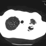
-
Image F. 14/5/99
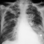
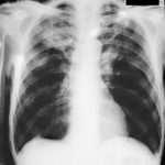
Compare with B, showing progressive enlargement of cavity and formation of fungal ball.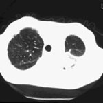
-
Image E 14/5/99
Showing enlargement of cavity at left apex and formation of a new cavity there.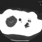
-
Image C. 30/3/99
Parenchymal or pleural disease adjacent to the mediasternum on the left with diffuse parenchymal disease. Also pleural based nodules bilaterally.


