Date: 1 December 2013
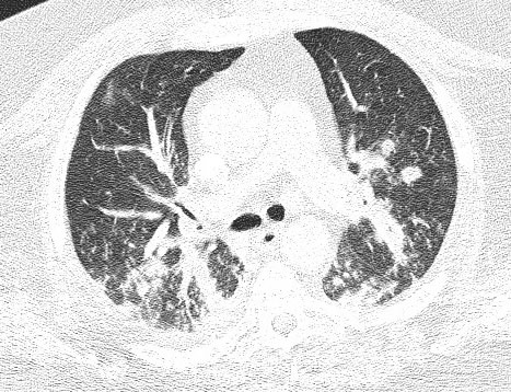
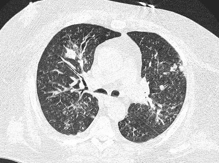
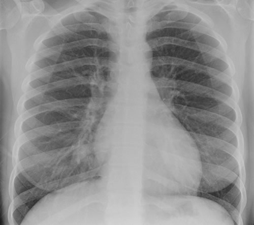
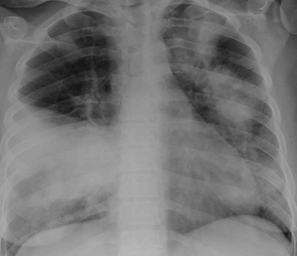
This patient is a 70 yr old, obese diabetic with aortic stenosis and COPD. He was admitted in early March 09, with collapse and loss of conciousness. His lungs appeared normal at this time – CT and X-ray 1. 10 days later he was admitted with increasing shortness of breath and chest X ray (F) showing widespread patchy consolidation. CT scan (B) showed bronchial dilatation, mucus plugging, nodular and bibasal consolidation. Multiple sputum samples grew Aspergillus fumigatus. The patient required intubation and remained in ITU for 160 days.
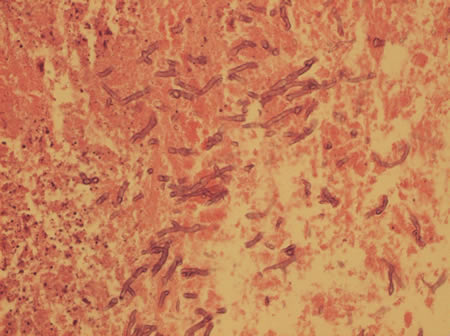
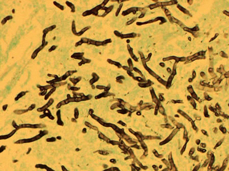
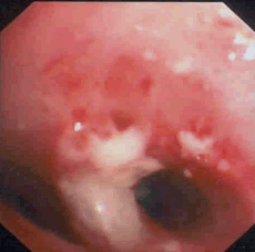
Bronchoscopy showed plaques in the major airways with more distal airways plugged with secretions resembling “cottage cheese”.There was severe contact bleeding and oedematous mucosa (I & J). Biopsy of the plaques showed fungal hyphae with a branching pattern consistent with aspergillus infection (G & H).
The patient was initially given IV and nebulised amphotericin B whilst on doses of hydrocortisone from 100-400 mg/day. Voriconazole was added with dose optimisation, and amphotericin discontinued. The patient improved gradually with voriconazole treatment over several months and for the latter month, gamma interferon was added into his regime which further improved his CT scan although some shadowing and bronchial wall thickening was still seen (D).
Copyright: n/a
Notes: n/a
Images library
-
Title
Legend
-
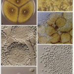
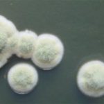
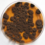
Pigmentation of Aspergillus versicolor colonies ranged from pale green to greenish-beige, pink-green, dark green and brown. Reverse is usually reddish. The growth rate is usually slow. Cultured on Sabouraud dextrose agar with chloramphenicol.
-
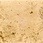
A Colonies on MEA after one week; B, C conidial heads with tip of conidiophire, x920; D conidial head, x 2330; E conidial heads x920
![aspvers[2] aspvers2](https://www.aspergillus.org.uk/wp-content/uploads/2017/10/aspvers2-150x150.jpg)
-
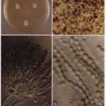
A Colonies on MEA + 20% sucrose after one week; B detail of colony showing columnar conidial heads x 44 ; C conidial heads x 920; D conidia x2330
-
Cultures are grown on malt extract agar for 5-7 days at 30°C.
Light microscopy-1000x stained with lacto-phenol and cotton blue.
-
A Colonies on MEA +20% sucrose after one week; B ascomata x 40; C conidiophores x 920; D ascospores x2330; E ascoma x 230; F portion of ascoma with asci and ascospores, x 920.
