Date: 26 November 2013
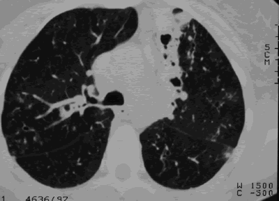
CT scans of thorax. Anterior left-sided bronchiectasis with extensive mucous plugging and with some proximal bronchiectasis and plugging on the right.
Copyright: n/a
Notes:
Extensive multilobar, varicose bronchiectasis, with some cyst formation most marked on the left anteriorly. Also some inhomogeneity of the pulmonary parenchyma secondary to air trapping in several affected segments.
Images library
-
Title
Legend
-
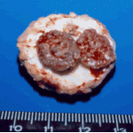
Gross pathology demonstrating the great pleural thickness and two cavities (upper lobe and superior segment of lower lobe) with fragments of fungal mass.

-
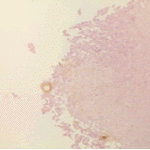
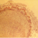
Histopathological appearance of a fungus ball. Note a conidial head resulting from fungal exposure to the air.
-
Histopathological appearance of a fungus ball caused by Scedosporium apiospermum. The presence of anneloconidia differentiates it from Aspergillus.

-
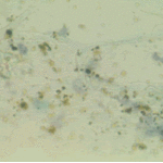
Chronic necrotising aspergillosis. Hyaline hyphal and calcium oxalate crystals obtained by needle aspirate biopsy from a diabetic patient with chronic necrotizing aspergillosis caused by Aspergillus niger (Papanicolaou, x 100).
-
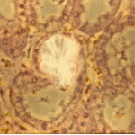
Aspergillus niger fungus ball and acute oxalosis. Higher magnification of adjacent replicate section.
-
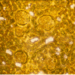
Oxalate crystals within renal tubuli (H&E, phase contrast, x 100). This patient developed acute oxalosis.
-
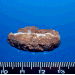
Lung surface. Fungus ball, severe parenchymal fibrosis and pleural thickening.

-
The periphery of the fungus ball is deeply eosinophilic because of the deposition of Splendore-Hoeppli material.

 ,
, 
 ,
,  ,
, 