Date: 1 November 2018
Copyright: n/a
Notes:
Professor Ken Haynes was a great fungal biologist with a keen eye for the Grand Vision, a loyal, supportive and hilarious friend to many in the fungal community, an inspiring mentor to innumerable junior scientists, and a loyal supporter of Fulham Football Club. He left us far too early, on 19th March this year at the age of 58, but he has entrusted us with a superb legacy in the field of molecular medical mycology.
Images library
-
Title
Legend
-
1 Axial computed tomography (CT) scans of the frontal sinus.
A: due to the long lasting pressure of mucus, the bone of the anterior wall of frontal sinus is thinned out and elevated anteriorly, forming a bulge. B: same situation as depicted in fig A: the posterior bony wall of frontal sinus is thinned out and extremely elevated posteriorly towards the frontal lobe of the brain. As depicted on the scan, a thin bony layer covering the dura could be recognized intraoperatively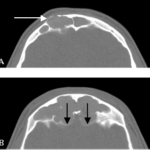
-
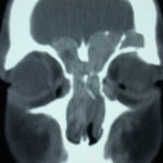
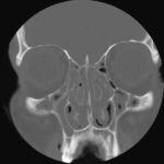
2 Same patient as 1 and 3, frontal CT
-
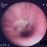
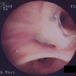
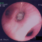
D. 6 months later, tenacious yellow secretions in L basal bronchial division
-
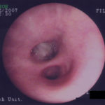
C. After suction the material was seen to extend distally – obstructing the right basal stem bronchus
-
B. After suction the material was seen to extend distally – obstructing the right basal stem bronchus
-
A. Necrotic mass prolapsing in and out of the distal right intermediate bronchus obscuring both the basal stem and basal division
