Date: 3 February 2014
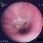
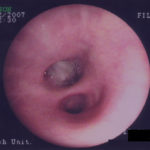
D. 6 months later, tenacious yellow secretions in L basal bronchial division
Copyright:
(© Fungal Research Trust)
Notes:
Initially it was incorrectly diagnosed as a bronchial carcinoma.
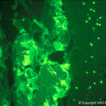
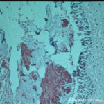
The material was allergic mucin with mucus and cellular debris arranged in a layered pattern. Cellular debris was almost entirely eosinophils with scattered Charcot-Leyden crystals. A Grocott stain showed multiple branching fungal hyphae, consistent with Aspergillus spp. Subsequently her total IgE rose to 750 KIU/L and Aspergillus specific RAST to 14.9 KUa/L.
Images library
-
Title
Legend
-
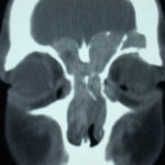
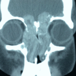
4 Total obstruction of the sinuses due to inflamed mucosa. (Patient 04)
-
1 Axial computed tomography (CT) scans of the frontal sinus.
A: due to the long lasting pressure of mucus, the bone of the anterior wall of frontal sinus is thinned out and elevated anteriorly, forming a bulge. B: same situation as depicted in fig A: the posterior bony wall of frontal sinus is thinned out and extremely elevated posteriorly towards the frontal lobe of the brain. As depicted on the scan, a thin bony layer covering the dura could be recognized intraoperatively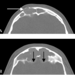
-
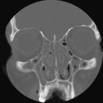
2 Same patient as 1 and 3, frontal CT
-
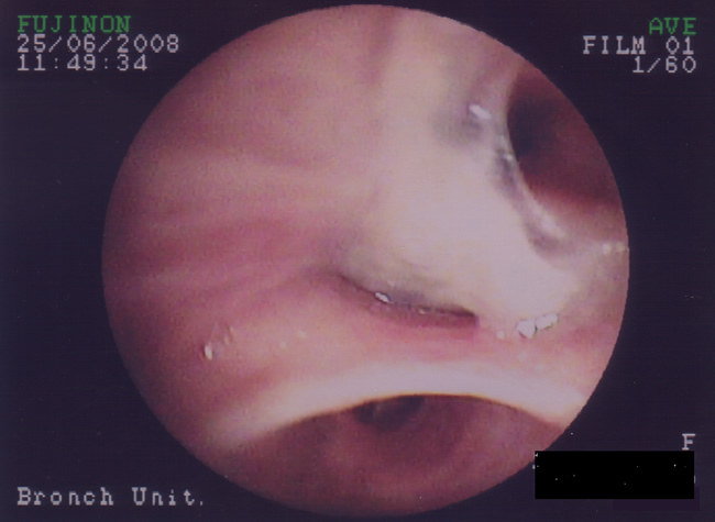
C. After suction the material was seen to extend distally – obstructing the right basal stem bronchus
-
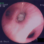
B. After suction the material was seen to extend distally – obstructing the right basal stem bronchus
-
A. Necrotic mass prolapsing in and out of the distal right intermediate bronchus obscuring both the basal stem and basal division
