Date: 6 August 2015
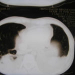
Percutaneous biopsy needle is seen vertically above the back
Copyright: n/a
Notes:
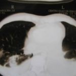
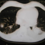
Under CT scan guidance, with the patient lying on their front, a percutaneous biopsy needle is seen vertically above the back, penetrating the skin, subcutaneous tissue and between 2 ribs. It is aimed at a an inflammatory area in the upper lobe of the lung, which defied diagnosis by other means. This area is much larger to access than the tiny nodule seen in in an identical location in the other lung. The lungs show considerable destruction of normal architecture, typical of emphysema with bullae, indicating that the patient was a heavy smoker.
Images library
-
Title
Legend
-
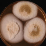
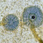
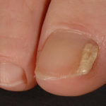
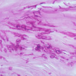
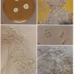
Image D & E. A case of onychomycosis associated with Aspergillus ochraceopetaliformis as described in Nail infection by Aspergillus ochraceopetaliformis. Med Mycol. 2009 Mar 9:1-5, 2009, Brasch J, Varga J, Jensen JM, Egberts F & Tintelnot K
 ,
,  ,
,  ,
,  ,
, 
-
Further details
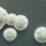
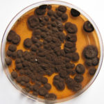
Image 5. Oral itraconazole pulse therapy was given to the patient (200 mg twice daily for 1 week, with 3 weeks off between successive pulses, for four pulses) and treatment was successful.
 ,
,  ,
,  ,
,  ,
, 
-
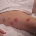
This patient was 28 yr old with adult lymphocytic leukaemia. She received induction chemotherapy and this infection developed 2 days after recovering from neutropenia.
 ,
,  ,
,  ,
,  ,
,  ,
,  ,
,  ,
,  ,
,  ,
, 
-
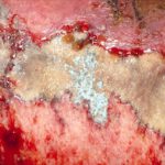
Close-up image of the lesion on the left thigh showing a mat of hyphae over the wound.
-
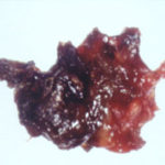
Eosinophilic mucin with A. flavus in the nasal cavity. Irregular crust of 2.5 cm from a patient diagnosed as allergic fungal sinusitis. Patient with allergic fungal sinusitis
-
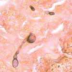
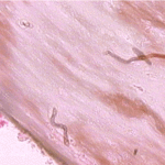
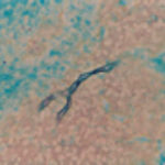
GMS stain of eosinophilic mucin reveals a darkly stained dichotomously branched A. flavus hyphae within cellular background. Patient with allergic fungal sinusitis
-
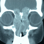
4 Total obstruction of the sinuses due to inflamed mucosa. (Patient 04)
