Date: 26 November 2013
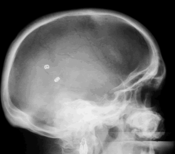
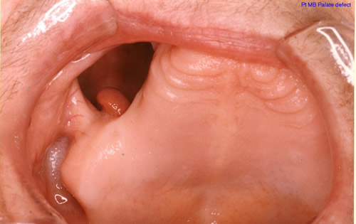
Patient MB X rays and CT scans. Chronic calcified maxillary sinusitis, patient had a palate defect.A. fumigatus cultured.
Images A&B Plain X rays antero-posterior and lateral, pre-operatively of Pt MB aged 76 who presented with unilateral nasal stuffiness and difficulty getting dentures fitted. She had hda these symptoms for many years. A large irregular calcified mass can be seen replacing the right maxillary sinus.
Images C D & E Coronal CT scan images of Pt MB showing a completely obstructed nasal cavity bilaterally and loss of internal nasal architecture. On the right side is large lamellar calcified lesion embedded in the extensive inflammatory material. Loss of bony margins is seen in numerous locations. This material was all removed surgically and showed mostly necrotic debris with Charcot-Leyden crystals and a few eosinophils and degenerate fungal hyphae. Aspergillus fumigatus was cultured from the material, especially infero-laterally on the right.
Image F Photograph through the mouth post-operatively showing the palate and a large defect in its right side. Through the defect can be seen the interior of the right maxillary sinus and nasal cavity with the inferior turbinate just visible.
Copyright: n/a
Notes: n/a
Images library
-
Title
Legend
-
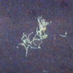
BAL specimen showing hyaline, septate hyphae consistent with Aspergillus, stained with Blankophor
-
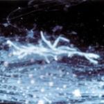
Mucous plug examined by light microscopy with KOH, showing a network of hyaline branching hyphae typical of Aspergillus, from a patient with ABPA.
-
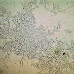
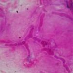
Corneal scraping stained with lactophenol cotton blue showing beaded septate hyphae not typical of either Fusarium spp or Aspergillus spp, being more consistent with a dematiceous (ie brown coloured) fungus
-
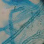
Corneal scrape with lactophenol cotton blue shows separate hyphae with Fusarium spp or Aspergillus spp.
-
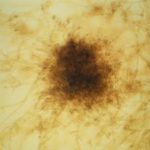
A filamentous fungus in the CSF of a patient with meningitis that grew Candida albicans in culture subsequently.
-
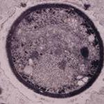
Transmission electron micrograph of a C. neoformans cell seen in CSF in an AIDS patients with remarkably little capsule present. These cells may be mistaken for lymphocytes.
-
India ink preparation of CSF showing multiple yeasts with large capsules, and narrow buds to smaller daughter cells, typical of C. neoformans
