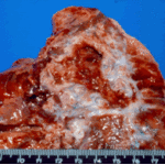
Date: 11 December 2013
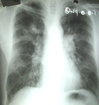
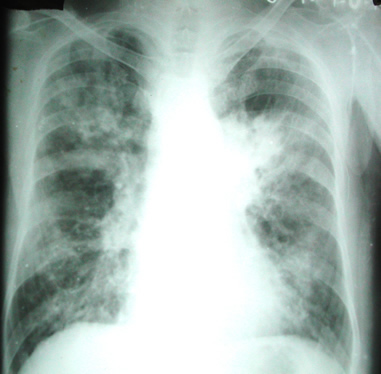
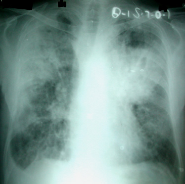
The chest X rays showed a rapid progression of lung disease- with bilateral upper zone and midzone consolidation and bilateral pleural effusion. Both lower lobes showed bronchiectasis in a central distribution along with centrilobular nodules and tree-in-bud pattern.
Copyright:
Case details kindly provided by Professor Arunaloke Chakrabarti Division of Mycology, Postgraduate Institute of Medical Education, Chandigarh, India
Notes: n/a
Images library
-
Title
Legend
-
Single fungal ball, moving. Radiographic appearance of a fungus ball, showing movement as the patient’s position changes.
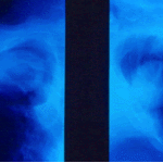
-
Oxalate crystals in the cavity wall surrounding an Aspergillus niger fungus ball (H&E, dark field, x 25).
-
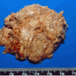
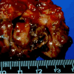
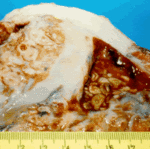
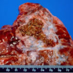
Aspergilloma patient. Gross pathology appearance of a fungus ball.
-
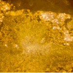
Conidiophores of Aspergillus fumigatus in the mass of the fungal ball surrounded by mycelia (H&E, x 400).
-
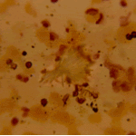
Aspergillus niger fungal ball. Calcium oxalate crystals in Aspergillus niger fungal ball. Also shown are darkly pigmented, rough-walled conidia associated with Aspergillus niger infection.
-
Aspergillus niger fungus ball within an old tuberculous cavern. This patient had diabetes, a disease commonly associated with A. niger infection.
-
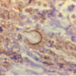
Conidial head and brown conidia in a section of a fungus ball caused by Aspergillus niger (H&E, x 400).



 ,
, 
 ,
, 