Date: 11 December 2013
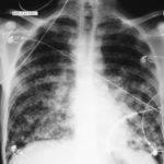
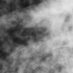
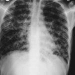
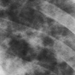
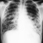
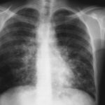
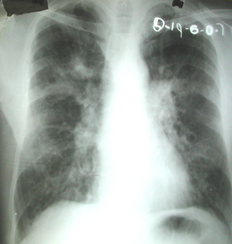
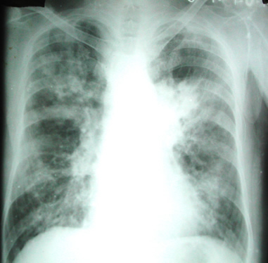
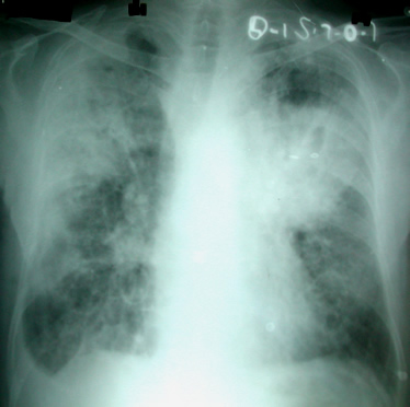
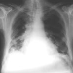
The chest X rays showed a rapid progression of lung disease- with bilateral upper zone and midzone consolidation and bilateral pleural effusion. Both lower lobes showed bronchiectasis in a central distribution along with centrilobular nodules and tree-in-bud pattern.
Copyright:
Case details kindly provided by Professor Arunaloke Chakrabarti Division of Mycology, Postgraduate Institute of Medical Education, Chandigarh, India
Notes: n/a
Images library
-
Title
Legend
-
Further details
Image 1. The chest x-ray shows extensive bilateral nodular disease, most consistent with a fungal infection, or possibly tuberculosis. He was treated with a bucket face mask with 80% oxygen and voriconazole.
 ,
,  ,
,  ,
,  ,
,  ,
, 
-
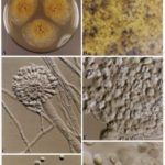
A Colonies on MEA +20 % sucrose after 2 weeks; B ascomata, x 40; C conidiophore of Aspergillus glaucus x 920;D conidiophore of Aspergillus glaucus x920 E. portion of ascoma with asci x 920. F ascospores x2330.
-
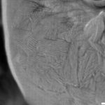
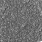
Scanning electron micrographs of A. fumigatus conidia of transformants rodB-02 (b). Size bar, 100 nm.

-
Scanning electron micrographs of A. fumigatus conidia of the wild-type G10 strain (a). Size bar, 100 nm.
-
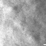
Scanning electron micrographs of A. fumigatus conidia of rodA rodB-26 (d).Size bar, 100 nm.
-
Scanning electron micrograph of an A.fumigatus conidium of rodA-47 (c), showing the hydrophobic rodlets covering the surface. Size bar, 100 nm.


 ,
, 
