Date: 26 November 2013
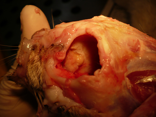
Nasal, sinus and orbital aspergillosis in a cat. The left nasal cavity and sinus were full of pus and debris and there was severe bone erosion from the nasal cavity into the rostromedial orbitthrough which pus was protruding
Copyright:
(Kindly provided by Martin L. Whitehead, BSc, PhD, BVSc, CertSAM, MRCVS & Peter W. Kettlewell, BVSc, MSc, MRCVS. Chipping Norton Veterinary Hospital, Albion Street, Chipping Norton, Oxon, OX7 5BN.)
Notes:
History : Nasal aspergillosis is relatively common in dogs but rare in cats. Our veterinary hospital in Oxfordshire was recently presented with a 13-year old female Burmilla cat with a history of left-side unilateral nasal discharge, a watery left eye with slight blepharospasm, occasional ‘twitching movements’ of the head, weight loss, inappetance and depression. Clinical examination was unremarkable except for left-side mucopurulent nasal discharge, left-side mild serous ocular discharge, and a soft subcutaneous swelling over the left frontal sinus. Haematology, blood biochemistry and urinalysis revealed diabetes mellitus but was otherwise unremarkable. Radiography under general anaesthesia revealed a diffuse soft tissue density within the left nasal cavity and left frontal sinus. Rhinoscopy revealed mucopurulent discharge on the left side but was otherwise unremarkable. Aspiration of the swelling over the left frontal sinus produced pus and this abscess was lanced and flushed. The frontal sinus was trephined and the sinus and nasal cavity flushed with saline. Tests for feline immunodeficiency virus and feline leukaemia virus and serology for Aspergillus were not carried out. The cat was started on insulin, ibafloxacin (Ibaflin, Intervet) and meloxicam (Metacam, Boehringer). Cytology of the material flushed from the frontal sinus and nasal cavity revealed fungal hyphae consistent with Aspergillus species and culture of this material yielded growth of a fungus which was morphologically similar to A. candidus (Awaiting molecular typing results). The cat was then started on oral itraconazole (Itrafungol, Janssen) 10 mg/kg p.o. SID. The abscess over the rostral frontal sinus did not heal and a second abscess appeared over the nasal bone just dorsal to the nose. Infusion of the frontal sinus and nasal cavity with topical antifungal medication was discussed with the owners, but as the cat was deteriorating they requested euthanasia. On post-mortem examination the right nasal cavity, frontal sinus and orbit were unaffected. The left nasal cavity and sinus were full of pus and debris and there was severe bone erosion from the nasal cavity into the rostromedial orbit through which pus was protruding. There was also severe bone erosion rostrally through the nasal bone and less severe bone erosion dorsally over the rostral part of the frontal sinus, these sites of bone erosion being at the location of the two subcutaneous abscesses.Feline nasal aspergillosis is extremely rare in the UK and to our knowledge this is the first reported case of orbital aspergillosis in the UK although nasal aspergillosis has been reported in other countries.
Images library
-
Title
Legend
-
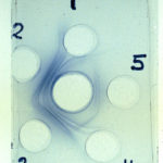
Aspergillus – Immunodiffusion Test showing stong immunoprecipitin lines against aspergillus.
-
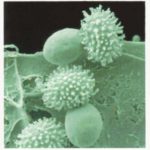
Talaromyces macrosporus- very short heat treatments of dormant ascospores at 85 C are able to activate these spores to germinate within 1 minute. One stage of germination is a very quick bursting of the thinly walled inner cell through the thick ornamented outer cell wall of the spore.
-
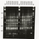
RAPD profiles of 14 A fumigatus isolates from Swedish and New Zealand saw mills, generated with primer R108. Mwt markers (lambda with Pst1) indicated by M.
-
These 3 images show non-union of the sternum post-aortic valve replacement as a result of local Aspergillus fumigatus infection. Underlying the sternum is some soft tissue which is presumptively also infection and in the most inferior image, the pericardium is also invoved.
The patient also is diabetic, with rhematoid arthritis and ulcerative colitis and occasionally receives a course of corticosteroids. The wound discharged pus which grew A. fumigatus. It was managed conservatively with itraconazole initally, with failure of therapy over 3 months.
 ,
,  ,
, 
-
CHEF gel of A. fumigatus chromosomes. The electrophoretic karyotype of two isolates of A. fumigatus is shown. The chromosomal bands have been resolved using Bio-Rad CHEF DRII equipment. The sizes of the bands vary between the two isolates and at least five bands have been resolved using the following conditions: chromosomal grade agarose (Bio-Rad) at 1.2 % in 1 x TAE buffer was used; the gel was run with a field strength of 1.8 V/cm for 48 h with a switch time of 2200 s and for 71 h with a switching ramp

-
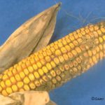
Aspergillus ear rot and storage mould – Aspergillus flavus and Aspergillus parasiticus can produce aflatoxins are generally known as storage fungi, but they can also cause ear rots in the field. These species are observed as a gray-green, powdery molds and they can be detected in corn because they produce compounds that are fluorescent under black light.
-
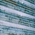
Environmental and sick building images. Dirty ventilation intake ducts
-
A.fumigatus conidium. Scanning electron micrograph of an A.fumigatus conidium showing the fascicles of hydrophobic rodlets covering the surface


