Date: 26 November 2013
Copyright: n/a
Notes: n/a
Images library
-
Title
Legend
-
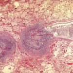
Medium power view of lung (H&E) in which there is invasion of lung parenchyma by hyphae characteristic of Aspergillus resulting in lung tissue infarction.
-
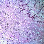
Vascular thrombosis. Medium power view (H&E) of a blood vessel occluded by fungal hyphae and thrombosis. Some fungal hyphae can be seen traversing the vessel wall.

-
Four colonies of Aspergillus on an agar plate containing rose bengal (to limit colony spending) and elastin fibres (light pink dots). Underneath and surrounding the colonies, the elastin fibres have gone, indicating enzymatic degradation

-
High power view of elastin fibres in an arterial wall being forced apart by Aspergillus hyphae. As Aspergillus is angiotropic and produces an elastase, it was uncertain how it traversed vessel walls. It appears to do so without any dissolution of elastin fibres.
-
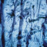
Medium power view (GMS) of hyphae seen within an arterial wall which is characteristic of angioinvasive Aspergillus.

-
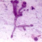
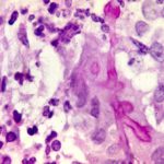
High power view (H&E) of a branching mould, consistent with Aspergillus inside a giant cell in a patient with chronic granulomatous disease.
-
High power view (GMS) of an Aspergillus hypha within a giant cell in a patient with chronic granulomatous disease infected with Aspergillus.

-
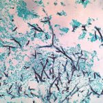
Medium power view (GMS) of the contents of a cerebral abscess in which there are hyphae typical of Aspergillus. Aspergillus fumigatus was grown from adjacent tissue.
-
Low power view (H&E) showing a pulmonary vein and a small bronchus infiltrated by fungal hyphae with associated necrotising inflammation.
