Date: 26 November 2013
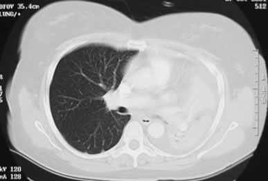
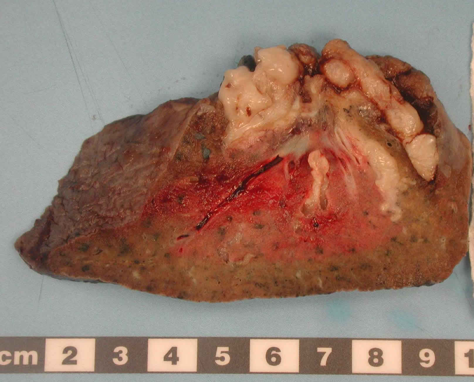
The patient underwent a pneumonectomy because of the severity of her disease process, and uncertainty about the diagnosis, prior to serology results being obtained.
Serology showed an IgE of 2600, with a strongly positive Aspergillus RAST test and weakly positive Aspergillus precipitins. Material removed at bronchoscopy showed eosinophilia. These features confirm a diagnosis of allergic bronchopulmonary aspergillosis (ABPA).
Copyright:
© Fungal Infection Trust
Notes:
Images library
-
Title
Legend
-
Image A
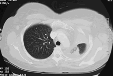
CT Scan 30/3/99
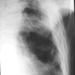
Showing extreme pleural thickening and 2 small cavities at apex of left lung.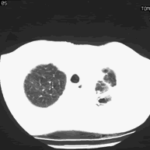
-
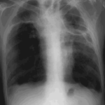
A 43 year old with smoking related emphysema was admitted to hospital with two separate episodes of haemoptysis. He had been in good health up to 1989, when he was diagnosed as having bilateral pulmonary tuberculosis. At that time a CT scan revealed a cavity in the left upper lobe (20.8cm2) with adjacent confluent infiltrates and pleural thickening. On bronchoscopic examination no abnormalities were noted and endobronchial biopsies did not reveal hyphae.
Over the next 4 years his condition deteriorated and a CT scan showed the left upper lobe cavity had increased to 40cm2. Itraconazole 400mg daily was prescribed. There was some clinical improvement on itraconazole but patient eventually deteriorated with breathlessness and with significant weight loss.
 ,
, 
-
Pt AR Interval development of chronic cavitary pulmonary aspergillosis in the context of sarcoidosis
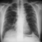
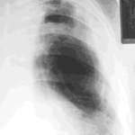
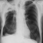
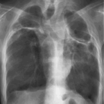
This patient was diagnosed with sarcoid after developing a chronic cough with the attached chest X-ray. In February 2003 the X-ray demonstrated bilateral extensive changes consistent with fibrocystic sarcoidosis with a complex cavitary area in both apices, more marked on the right. She was given a course of corticosteroids.
