Date: 26 November 2013
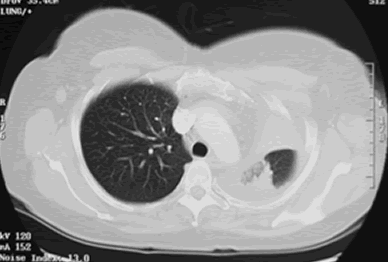
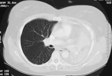
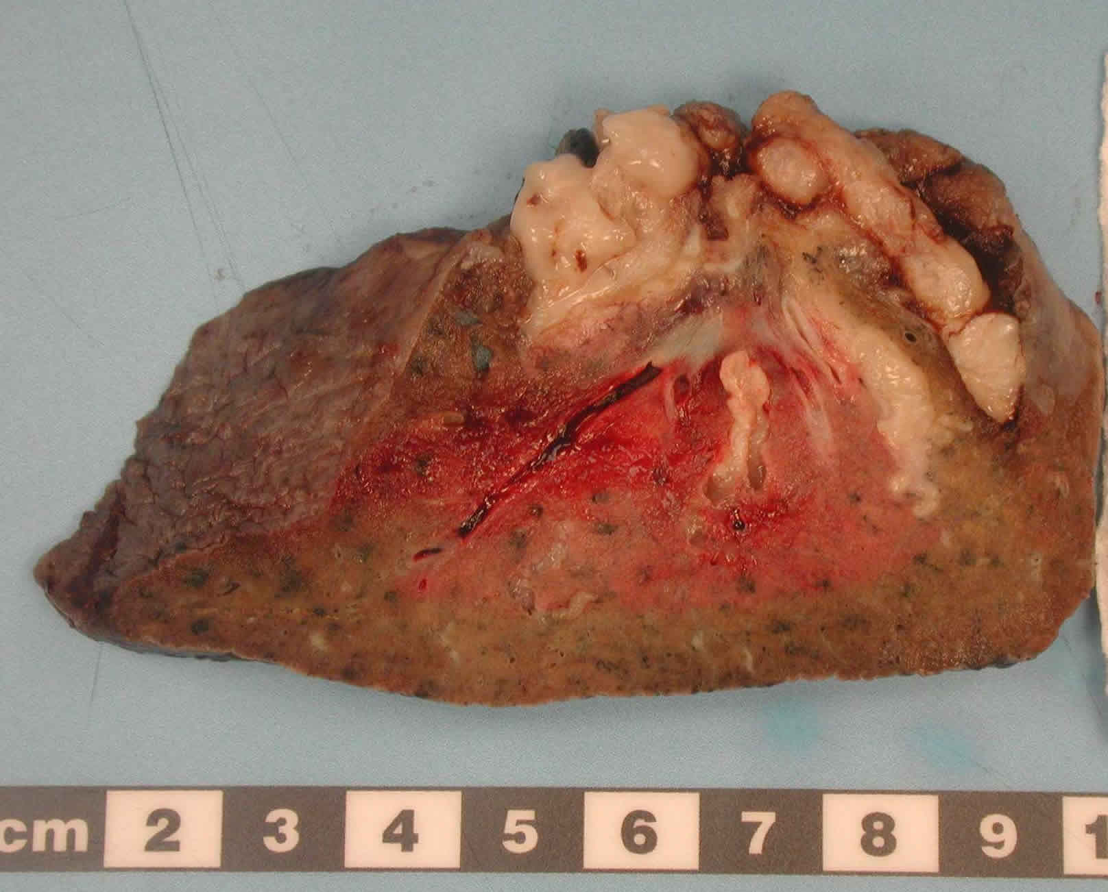
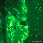
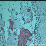
The patient underwent a pneumonectomy because of the severity of her disease process, and uncertainty about the diagnosis, prior to serology results being obtained.
Serology showed an IgE of 2600, with a strongly positive Aspergillus RAST test and weakly positive Aspergillus precipitins. Material removed at bronchoscopy showed eosinophilia. These features confirm a diagnosis of allergic bronchopulmonary aspergillosis (ABPA).
Copyright:
© Fungal Infection Trust
Notes:
Images library
-
Title
Legend
-
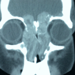
4 Total obstruction of the sinuses due to inflamed mucosa. (Patient 04)
-
1 Axial computed tomography (CT) scans of the frontal sinus.
A: due to the long lasting pressure of mucus, the bone of the anterior wall of frontal sinus is thinned out and elevated anteriorly, forming a bulge. B: same situation as depicted in fig A: the posterior bony wall of frontal sinus is thinned out and extremely elevated posteriorly towards the frontal lobe of the brain. As depicted on the scan, a thin bony layer covering the dura could be recognized intraoperatively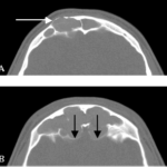
-
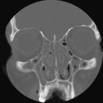
2 Same patient as 1 and 3, frontal CT
-
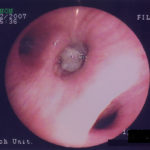
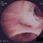
D. 6 months later, tenacious yellow secretions in L basal bronchial division
-
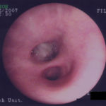
C. After suction the material was seen to extend distally – obstructing the right basal stem bronchus
-
B. After suction the material was seen to extend distally – obstructing the right basal stem bronchus
