Date: 21 January 2014
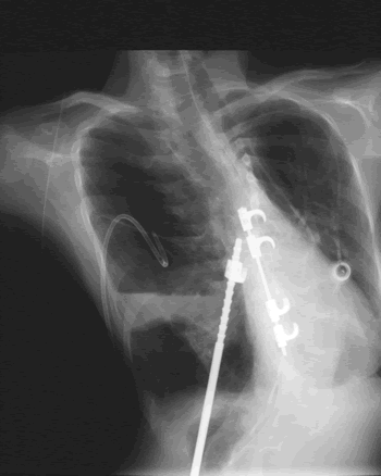
The chest is distorted by a deformity of the back and ribs.
Copyright: n/a
Notes:
This patient’s X-ray is complex. The chest is distorted by a deformity of the back and ribs. Substantial metalwork following a spinal fusion is in place to support the vertebral column and part of this overlies the heart and part of it crosses the left lung. The patient also has a portacath device in-situ over the right lung, which allows i.v. antibiotics to be given. A needle is in-situ inside the portacath device. An external drainage tube is currently in-situ in a large air cavity and left upper thorax. This cavity contains mostly air but there is some fluid with the fluid level at its base. Underneath this large pyopneumothorax is a normal component of left lower lobe. The heart is very substantially moved to the right of the lung because of a previous right lower lobe resection. There is no evidence of aspergillosis on this x-ray as it stands.
Images library
-
Title
Legend
-
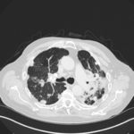
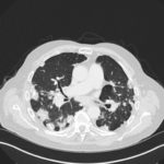
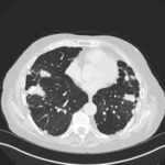
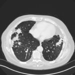
Mr RM is 80 and an ex-coal miner.He developed pneumoconiosis from exposure to coal dust. He also developed rheumatoid arthritis and the combination of this disease and pneumoconiosis is called Caplan’s syndrome.
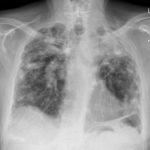
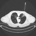
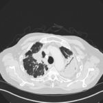
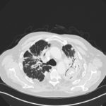
His chest Xray in early 2015 shows extensive bilateral pulmonary shadowing with solid looking nodules superimposed on abnormal lung fields, contraction of his left lung with an elevated diaphragm and a large left upper lobe aspergilloma, displaying a classic air crescent. His CT scan from mid 2014 demonstrates a large aspergilloma in a cavity on the left, with marked pleural thickening around it, which is partially ‘calcified’ towards its base. Inferiorly on other images,remarkable pleural thickening and fibrotic irregular and spiculated nodules are seen, most partially calcified.
 ,
,  ,
,  ,
,  ,
,  ,
,  ,
, 