Date: 6 November 2014
Copyright: n/a
Notes:
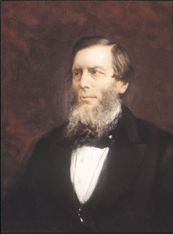
Bennett’s portrait at the Royal College of Physicians of Edinburgh
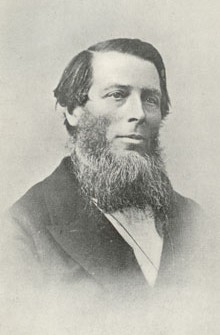
In his 1842 paper Bennett gave the earliest description of pulmonary aspergillosis. Bennett was one of the first to recognise the importance of the microscope in the clinical investigation of disease and his use of the instrument was central to identifying the presence of a fungus in the sputum and, post mortem, lungs of the patient with aspergillosis.
A biography on Wikipedia
An obituary from the British Medical Journal of 1875
Images library
-
Title
Legend
-
1 Axial computed tomography (CT) scans of the frontal sinus.
A: due to the long lasting pressure of mucus, the bone of the anterior wall of frontal sinus is thinned out and elevated anteriorly, forming a bulge. B: same situation as depicted in fig A: the posterior bony wall of frontal sinus is thinned out and extremely elevated posteriorly towards the frontal lobe of the brain. As depicted on the scan, a thin bony layer covering the dura could be recognized intraoperatively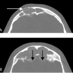
-
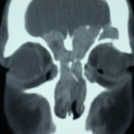
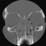
2 Same patient as 1 and 3, frontal CT
-
D. 6 months later, tenacious yellow secretions in L basal bronchial division
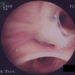
-
C. After suction the material was seen to extend distally – obstructing the right basal stem bronchus
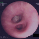
-
B. After suction the material was seen to extend distally – obstructing the right basal stem bronchus
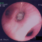
-
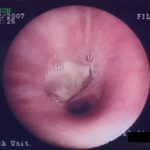
A. Necrotic mass prolapsing in and out of the distal right intermediate bronchus obscuring both the basal stem and basal division
