Date: 26 November 2013
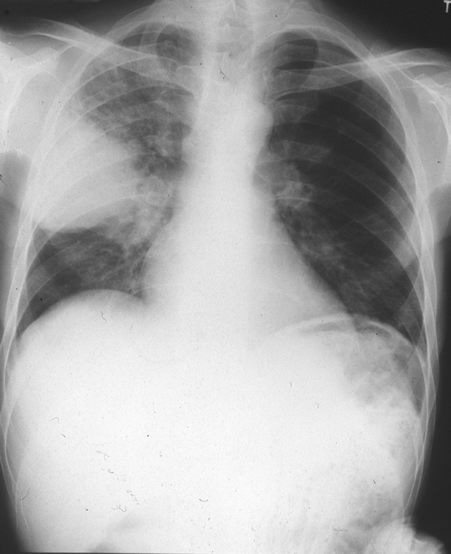
Chest X ray after 4 days, prior to treatment, showing massive increase in volume of lesion (Fig 2)
Copyright: n/a
Notes:
His case has been previously reported (Denning DW, Williams AH). Invasive pulmonary aspergillosis diagnosed by blood culture and successfully treated. Br J Dis Chest (1987) 81, 300).
Chest X ray after 4 days, prior to treatment, showing massive increase in volume of lesion. He started amphotericin B and flucytosineb that day and responded over 10 weeks.
Images library
-
Title
Legend
-
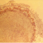
The periphery of the fungus ball is deeply eosinophilic because of the deposition of Splendore-Hoeppli material.
-
Single fungal ball, moving. Radiographic appearance of a fungus ball, showing movement as the patient’s position changes.
-
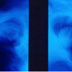
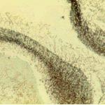
Oxalate crystals in the cavity wall surrounding an Aspergillus niger fungus ball (H&E, dark field, x 25).
-
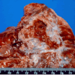
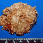
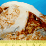
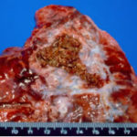
Aspergilloma patient. Gross pathology appearance of a fungus ball.
-
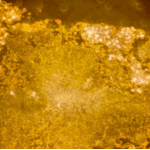
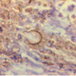
Conidiophores of Aspergillus fumigatus in the mass of the fungal ball surrounded by mycelia (H&E, x 400).
-
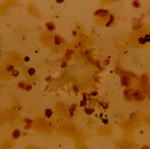
Aspergillus niger fungal ball. Calcium oxalate crystals in Aspergillus niger fungal ball. Also shown are darkly pigmented, rough-walled conidia associated with Aspergillus niger infection.
-
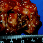
Aspergillus niger fungus ball within an old tuberculous cavern. This patient had diabetes, a disease commonly associated with A. niger infection.

 ,
, 
 ,
, 