Date: 26 November 2013
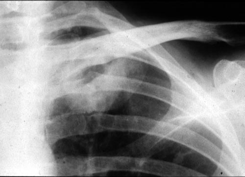
IPA in late stage AIDS, pt TB
Copyright: n/a
Notes:
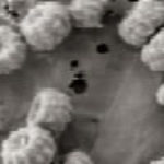
Close up of cavity of left upper-lobe, proven to be invasive pulmonary aspergillosis at autopsy.
Pulmonary aspergillosis in a patient who had AIDS for 3 years (CD4 count. <10/mm3). Right middle-lobe consolidation and a left upper-lobe cavity as seen in a previously normal lung; the diagnosis was made by culture of a bronchoalveolar lavage specimen and was confirmed at autopsy. (This was published in (Khoo S, Denning DW. Aspergillus infection in the acquired immune deficiency syndrome. Clin Infect Dis 1994; 19 (suppl 1): S41-48.)
Images library
-
Title
Legend
-
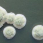
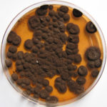
Pigmentation of Aspergillus versicolor colonies ranged from pale green to greenish-beige, pink-green, dark green and brown. Reverse is usually reddish. The growth rate is usually slow. Cultured on Sabouraud dextrose agar with chloramphenicol.
-
A Colonies on MEA after one week; B, C conidial heads with tip of conidiophire, x920; D conidial head, x 2330; E conidial heads x920
![aspvers[2] aspvers2](https://www.aspergillus.org.uk/wp-content/uploads/2017/10/aspvers2-150x150.jpg)
-
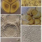
A Colonies on MEA + 20% sucrose after one week; B detail of colony showing columnar conidial heads x 44 ; C conidial heads x 920; D conidia x2330
-
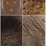
Cultures are grown on malt extract agar for 5-7 days at 30°C.
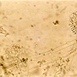
Light microscopy-1000x stained with lacto-phenol and cotton blue.
-
A Colonies on MEA +20% sucrose after one week; B ascomata x 40; C conidiophores x 920; D ascospores x2330; E ascoma x 230; F portion of ascoma with asci and ascospores, x 920.
