Date: 26 November 2013
IPA in late stage AIDS, pt TB
Copyright: n/a
Notes:
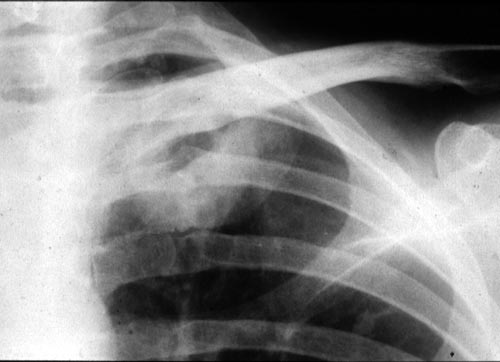
Close up of cavity of left upper-lobe, proven to be invasive pulmonary aspergillosis at autopsy.
Pulmonary aspergillosis in a patient who had AIDS for 3 years (CD4 count. <10/mm3). Right middle-lobe consolidation and a left upper-lobe cavity as seen in a previously normal lung; the diagnosis was made by culture of a bronchoalveolar lavage specimen and was confirmed at autopsy. (This was published in (Khoo S, Denning DW. Aspergillus infection in the acquired immune deficiency syndrome. Clin Infect Dis 1994; 19 (suppl 1): S41-48.)
Images library
-
Title
Legend
-
Necrotic lung tissue in culture
Af=Colony of Aspergillus fumigatus
B=bacterial colonies
L=lung tissue
-
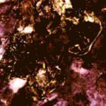
A photograph of part of the upper lung lobe of an immunosuppressed patient. The lung tissue shows extenive areas of necrosis due to invasive colonisation.
-
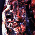
A photograph of a cavity in the upper lobe of the lung of a patient with ankylosing spondylitis. Such cavitation,which may be confused with prior tuberculosis, can follow fibrosis.
-
Plugs stained with Methenamine/silverActively growing mycelia of the fungus are a deep brown/black. Counterstaining shows the dense mucus of the plugs as predominantly orange.
-
Sputum from an asthmatic patient showing plugs(casts). The development of plugs coincided with an increased prevalence and severity of episodes of asthma

-
microscopic characters Conidiophore stipes(C)1300-2800um long:Vesicles(V)40-70um wide,clavate:Phialides(Ph) uniseriate:Conidia(Con)3.5-4.0um long,smooth walled.

-
microscopic characters Conidiophore stipes(C)225-350um arising from hyphae(Hy):Vesicles(Ves)15-25um wide:Phialides(Ph)uniseriate:Conidia(Con)2.4-3.0um spherical to ovoid,roughened.


