Date: 3 April 2014
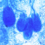
PAS stain. An example of Aspergillus fumigatus.
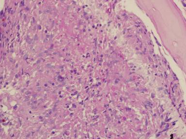
(PAS-stained) in a patient with chronic granulomatous disease showing a 45 degree branching hypha within a giant cell. Rather bulbous hyphal ends are also seem, which is sometimes found inAspergillus spp. infections, histologically. (x800)
Copyright: n/a
Notes:
Comparison of GMS and PAS stains. Patient with disseminated Trichosporon spp. infection. Both x60. In the GMS image, substantial background staining of elastin is seen, with more prominent yeasts superimposed. In contrast, the PAS stain shows the tissue morphology, with bright pink yeasts also visible.
Images library
-
Title
Legend
-
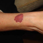
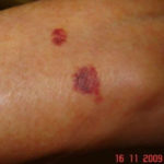
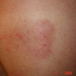
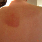
Drug rashes: Drug interactions between steroids and anti-fungal drugs – (ecchymosis)
 ,
,  ,
,  ,
, 
-
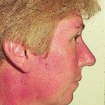
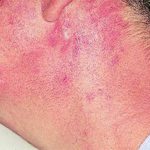
Reference: Muco-cutaneous retinoid effects and facial erythema related to the novel triazole antifungal agent voriconazole. Denning, DW & Griffiths, CEM. Clin.Exp Dermatol 2001, 26(8), 648-53.
Courtesy of Dr D Denning, Wythenshawe Hospital, Manchester.(© Fungal Research Trust)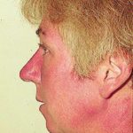 ,
,  ,
, 
-
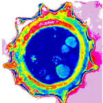
Micrographs of A. niger conidia & conidial heads provided by Amaliya Stepanova, Head of Laboratory pathomorphology and cytology at Kashkin Research Institute, Russian Federation.
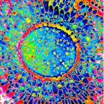 ,
, 
-
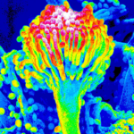
Micrographs of A. terreus conidia & conidial heads provided by Amaliya Stepanova, , Head of Laboratory pathomorphology and cytology at Kashkin Research Institute, Russian Federation.
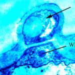 ,
,  ,
, 
-
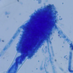
Micrographs of A. fumigatus conidia & conidial heads provided by Amaliya Stepanova, , Head of Laboratory pathomorphology and cytology at Kashkin Research Institute, Russian Federation.
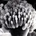 ,
, 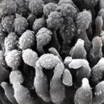 ,
, 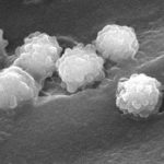 ,
,  ,
, 
-
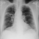
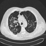
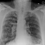
Patients has history of ABPA complicating long standing asthma. His total IgE has fluctuated between 2,200 and 4,600 KU/L, his Aspergillus IgE between 36.3 and 65.4 kAU/L and Aspergillus IgG from 87-154 mg/L. He has been taking long term itraconazole.
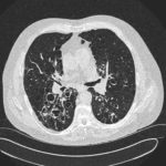 ,
,  ,
,  ,
, 

 ,
,  ,
, 