Date: 3 April 2014
PAS stain. An example of Aspergillus fumigatus.
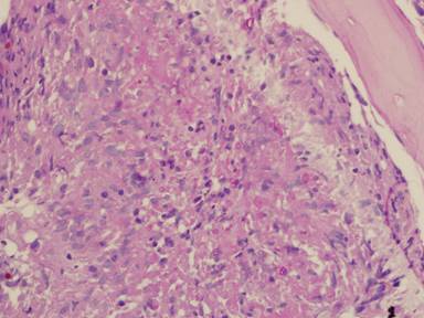
(PAS-stained) in a patient with chronic granulomatous disease showing a 45 degree branching hypha within a giant cell. Rather bulbous hyphal ends are also seem, which is sometimes found inAspergillus spp. infections, histologically. (x800)
Copyright: n/a
Notes:
Comparison of GMS and PAS stains. Patient with disseminated Trichosporon spp. infection. Both x60. In the GMS image, substantial background staining of elastin is seen, with more prominent yeasts superimposed. In contrast, the PAS stain shows the tissue morphology, with bright pink yeasts also visible.
Images library
-
Title
Legend
-
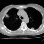
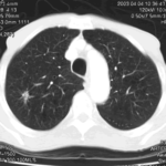
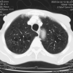
Nodules and areas of atelectasis are seen at both bases. He later died.

-
Further details
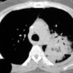
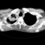
It is clearly a relatively small cavitary lesion, and the patient was almost asymptomatic. This response was a ‘stable’ response. The patient was included in the report Denning DW, Lee JY, Hostetler JS, Pappas P, Kauffman CA, Dewsnup DH, Galgiani JN, Graybill JR, Sugar AM, Catanzaro A, Gallis H, Perfect JR, Dockery B, Dismukes WE, Stevens DA, NIAID Mycoses Study Group multicenter trial of oral itraconazole therapy of invasive aspergillosis. Am J Med 1994; 97: 135-144.
 ,
, 
-
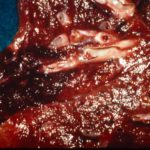
Well demarcated pulmonary infarction is well seen in this close-up of the lung at autopsy in a patient with histologically confirmed invasive aspergillosis. Angio invasion is characteristic of invasive aspergillosis, is associated with a worse prognosis, but is not always seen.
-
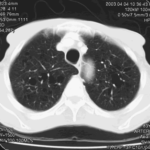
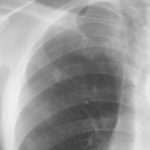
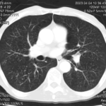
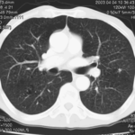
This 83 year old man presented with weight loss to a lung cancer clinic in mid 2003.
 ,
,  ,
,  ,
,  ,
,  ,
, 