Date: 3 April 2014
PAS stain. An example of Aspergillus fumigatus.
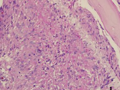
(PAS-stained) in a patient with chronic granulomatous disease showing a 45 degree branching hypha within a giant cell. Rather bulbous hyphal ends are also seem, which is sometimes found inAspergillus spp. infections, histologically. (x800)
Copyright: n/a
Notes:
Comparison of GMS and PAS stains. Patient with disseminated Trichosporon spp. infection. Both x60. In the GMS image, substantial background staining of elastin is seen, with more prominent yeasts superimposed. In contrast, the PAS stain shows the tissue morphology, with bright pink yeasts also visible.
Images library
-
Title
Legend
-
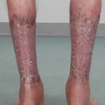
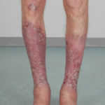
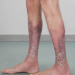
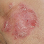
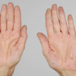
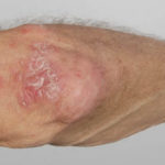
After 3 weeks of posaconazole given for chronic pulmonary aspergillosis, patient NC had a remarkable exacerbation of psoriasis. He had had psoriasis for years, with little trouble and almost no treatment. After taking posaconazole 400mg twice daily, he developed psoriatic plaques on his hands for the first time ever. The plaques on his lower legs became confluent. This occurred in association with worsening chest symptoms, notably increased coughing, more breathlessness and increasing oxygen requirement.
Posaconazole was stopped after 3 weeks, and 2 weeks later he was still very symptomatic with his chest. This responded to a 2 week course of corticosteroids, and his psoriasis also improved.
 ,
,  ,
,  ,
,  ,
,  ,
, 
-
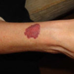
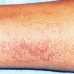
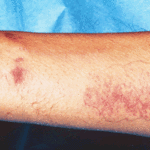
Patient PC: An example of localised caspofungin rash and phlebitis related to caspofungin infusion.
 ,
, 
-
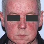
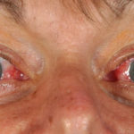
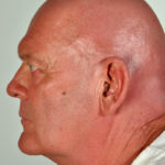
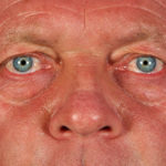
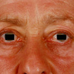
This 55 year old man with asthma, ABPA, severe bronchiectasis and lung fibrosis was treated with voriconazole, starting in June 2010. He had developed increasing dyspnoea on itraconazole for over 7 years, and his total IgE remained at 1100 KIU/L. He had marked photopsia (visual hallucinations) and facial erythema in the first 3 weeks of therapy. His trough voriconazole concentration was 1.17 mg/L. Over 3 months, he had minor improvement in his breathlessness but continued facial erythema, despite factor 50 sunblock. After 5 months of therapy his facial rash has altered to show acneiform lesions with localised crusting and background severe erythema. His face effectively crusted over, and he stopped therapy.
Over the next 3 weeks his facial appearance slowly improved .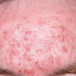 ,
,  ,
, 