Date: 3 April 2014
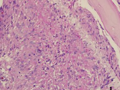
PAS stain. An example of Aspergillus fumigatus.
(PAS-stained) in a patient with chronic granulomatous disease showing a 45 degree branching hypha within a giant cell. Rather bulbous hyphal ends are also seem, which is sometimes found inAspergillus spp. infections, histologically. (x800)
Copyright: n/a
Notes:
Comparison of GMS and PAS stains. Patient with disseminated Trichosporon spp. infection. Both x60. In the GMS image, substantial background staining of elastin is seen, with more prominent yeasts superimposed. In contrast, the PAS stain shows the tissue morphology, with bright pink yeasts also visible.
Images library
-
Title
Legend
-
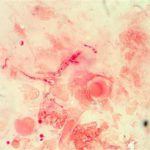
Corneal ulcer – gram stain. Corneal scrapings were taken from a 67 yr old farmer presenting with a corneal ulcer of the right eye. A piece of vegetable matter was embedded in the cornea and scrapings were done. Gram stain (500x magnification) showed numerous septate hyphae. Cultures grew a small amount of A fumigatus.

-
Aspergillus keratitis. Central lesion in aspergillus keratitis following a corneal foreign body which made a good response to topical treatment alone, albeit over 2 months intensive treatment.
-
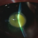
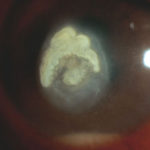
Aspergillus keratitis. B- Severe central aspergillus infection with a “cheesey†looking area of the lesion and hypopyon (fluid level of inflammatory cells in the anterior chamber)
-
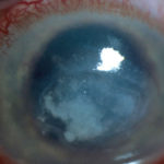
Aspergillus keratitis. A- Severe aspergillus infection with large area of corneal ulceration and deep stromal involvement
-
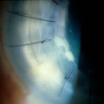
Candida keratitis. Focal candida keratitis as an unusual cause of a suture related infection following corneal transplantation for non infective indication
-
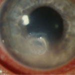
Candida keratitis. Subacute onset of candida keratitis in a young adult in whom dust blew into her eye in Greece. A slightly “feathery†edge to stromal involvement is suggestive of fungal infection
-
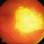
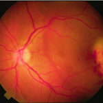
Aspergillus endopthalmitis. Temporal necrosis due to Aspergillus endopthalmitis as part of disseminated disease. No evidence of vitritis. Systemic treatment essential.
-
Aspergillus endopthalmitis. Large scarred area of the choroid following healing after Aspergillus endopthalmitis
