Date: 26 November 2013
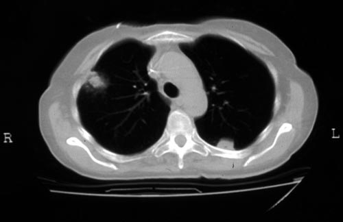
Halo sign in IPA
Copyright: n/a
Notes:
CT scan of a neutropenia patient with leukaemia who has 2 lesions. One, on the right, is nodular, abuts on the pleura and is surrounded by a (grey) low attenuation area, referred to as the “halo” sign. This is virtually only seen in invasive fungal infections of the lung, especially early in the course of the disease, during neutropenia. The other lesion visible on this scan, posteriorly on the left, is also typical of invasive pulmonary aspergillosis in that it is pleura-based and has sharply angulated sides typical of vascular invasion and infarction of small lung segments. There is the suggestion of a “halo” sign anteriorly, but there is less confidence in this appearance (compared with the other) because it is only on one side of the lesion.
Images library
-
Title
Legend
-
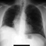
The chest x-ray shows a patient who had a left lung transplanted in May 2003 for cryptogenic fibrosing alveolitis, which was diagnosed post-transplant as sarcoidosis.
-
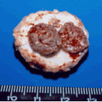
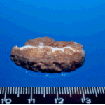
Gross pathology demonstrating the great pleural thickness and two cavities (upper lobe and superior segment of lower lobe) with fragments of fungal mass.

-
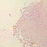
Histopathological appearance of a fungus ball. Note a conidial head resulting from fungal exposure to the air.
-
Histopathological appearance of a fungus ball caused by Scedosporium apiospermum. The presence of anneloconidia differentiates it from Aspergillus.

-
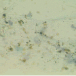
Chronic necrotising aspergillosis. Hyaline hyphal and calcium oxalate crystals obtained by needle aspirate biopsy from a diabetic patient with chronic necrotizing aspergillosis caused by Aspergillus niger (Papanicolaou, x 100).
-
Aspergillus niger fungus ball and acute oxalosis. Higher magnification of adjacent replicate section.
-
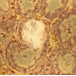
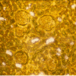
Oxalate crystals within renal tubuli (H&E, phase contrast, x 100). This patient developed acute oxalosis.
-
Lung surface. Fungus ball, severe parenchymal fibrosis and pleural thickening.


 ,
, 
 ,
,  ,
, 