Date: 26 November 2013
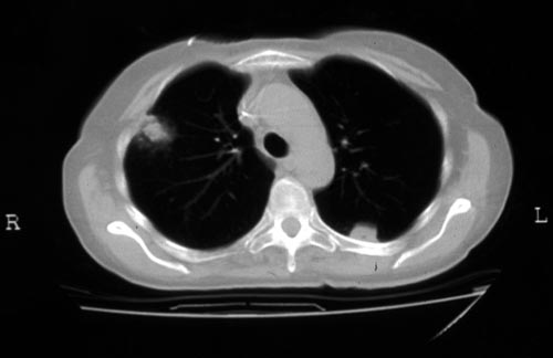
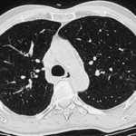
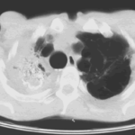
Halo sign in IPA
Copyright: n/a
Notes:
CT scan of a neutropenia patient with leukaemia who has 2 lesions. One, on the right, is nodular, abuts on the pleura and is surrounded by a (grey) low attenuation area, referred to as the “halo” sign. This is virtually only seen in invasive fungal infections of the lung, especially early in the course of the disease, during neutropenia. The other lesion visible on this scan, posteriorly on the left, is also typical of invasive pulmonary aspergillosis in that it is pleura-based and has sharply angulated sides typical of vascular invasion and infarction of small lung segments. There is the suggestion of a “halo” sign anteriorly, but there is less confidence in this appearance (compared with the other) because it is only on one side of the lesion.
Images library
-
Title
Legend
-
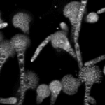
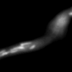
Mitochondria organisation: GFP fluorescence micrographs showing mitochondrial organisation in an A.nidulans strain with GFP mitochondria, grown at 25°C in minimal media.
-
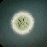
Colony morphology of A.nidulans SRF200 after two days at 37°C
-
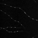
Aspergillus nidulans. Cell nuclei-Ds red. DsRed fluorescence micrographs showing nuclear distribution in an A.nidulans germling with dsRed stained nuclei

-
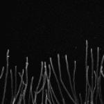
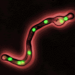
Cell Biology – Aspergillus nidulans. Cell nuclei-GFP. Nuclear distribution: GFP fluorescence mirographs showing fungal cell morphology and nuclear distribution in A.nidulans. GFP stained nuclei,grown at 25°C in minimal media O/N
-
High resolution CT scan of chest.CT scan demonstrating remarkable bronchial wall thickening of the right main bronchus and main branches, in context of longstanding ABPA


