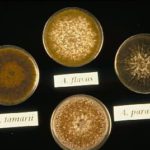
Date: 26 November 2013
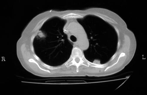
Halo sign in IPA
Copyright: n/a
Notes:
CT scan of a neutropenia patient with leukaemia who has 2 lesions. One, on the right, is nodular, abuts on the pleura and is surrounded by a (grey) low attenuation area, referred to as the “halo” sign. This is virtually only seen in invasive fungal infections of the lung, especially early in the course of the disease, during neutropenia. The other lesion visible on this scan, posteriorly on the left, is also typical of invasive pulmonary aspergillosis in that it is pleura-based and has sharply angulated sides typical of vascular invasion and infarction of small lung segments. There is the suggestion of a “halo” sign anteriorly, but there is less confidence in this appearance (compared with the other) because it is only on one side of the lesion.
Images library
-
Title
Legend
-
Canker: Grapevine. A. niger canker on Crimson Seedless grapes
-
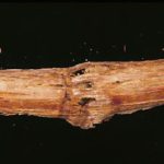
Canker: Grapevine. Canker caused by A. niger on trunk of Red Globe grapes

-
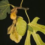
Blight: Fig leaf. Shoot and leaf blight caused by infection of fig fruit by A. niger
-
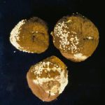
Fresh fruit: Plum. Infection of Friar plum with A. niger, showing many white sclerotia
-
Infection of a fresh Elegant Lady peach by A.niger

-
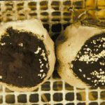
Fresh fruit: Nectarine. A. niger infected Fantasia nectarines showing abundance of white sclerotia
-
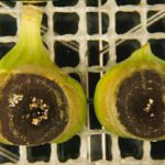
Fresh fruit: Fig. Sclerotia of A. niger on Calimyrna figs 6 days after inoculation
-
Fresh fruit: Fig. Infection of fresh fig by A. niger (early infection)

-
Fresh fruit: Fig. A. parasiticus infection on Calimyrna fig


