Date: 26 November 2013
Copyright: n/a
Notes: n/a
Images library
-
Title
Legend
-
Further details
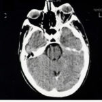
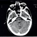
Image A. The bone appears normal, but the features are most consistent with infection, less so with a pseudotumour. Bone windows not shown. Biopsy demonstrated hyphal invasion and cultures grew A. fumigatus.
 ,
,  ,
,  ,
, 
-
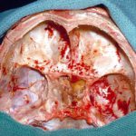
Bilateral Aspergillus osteomyelitis of the base of the skull in a non-immunocompromised woman, aged 38, following Aspergillus sinusitis. This was eventually cured with an 18 month course of voriconazole.
-
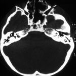
In this non-immunocompromised patient with histologically and culture proven invasive aspergillosis most of the landmarks have been surgically removed during 3 operations.

-
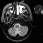
The maxillary sinus on the left is completely opacified with high signal, The high signal seen in the maxillary sinus on the right is of marginal significance. The sinuses and other cerebellum are normal.
-
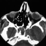
CT scan of the sinuses shows a completely opacified sphenoid sinus in this man with AIDS (Khoo S, Denning DW. Invasive aspergillosis in patients with AIDS Clin Infect Dis 1994; 19 (suppl 1): S41-8.) He had complained of headache for about 3 months before this scan was done. In addition to sinus disease, erosion of disease through the left lateral wall of the sinus into the arterior horn of the temporal lobe, can be seen leading to a cerebral abscess. The patient was treated with itraconazole, but had undetectable serum concentrations because of prior phenytoin, and died after nine days of therapy of an MRSA bacteraemia. He was reported as an “other failure” in Denning DW, Lee JY, Hostetler JS, Pappas P, Kauffman CA, Dewsnup DH, Galgiani JN, Graybill JR, Sugar AM, Catanzaro A, Gallis H, Perfect JR, Dockery B, Dismukes WE, Stevens DA, NIAID Mycoses Study Group multicenter trial of oral itraconazole therapy of invasive aspergillosis. Am J Med 1994; 97: 135-144.
Cultures from the sinus and brain abcess grew A.fumigatus.
 ,
, 