Date: 26 November 2013
Copyright: n/a
Notes: n/a
Images library
-
Title
Legend
-
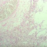
Pulmonary aspergillosis (PAS) (parrot C). Section of lung from parrot C stained by periodic acid schiff (PAS) demonstrating the hyphal material. Aspergillus spp and Bacillus cereus were cultured from the lesions.

-
Pulmonary aspergillosis (K&E) (parrot C). Tissue from an individually housed and recently purchased, 6 month old African grey parrot found dead in the cage. Necropsy examination revealed focal necrosis of the left lung. This section stained by haematoxylin and eosin reveals septate fungal hyphae within the lung parenchyma. Similar hyphae were located in the walls and lumen of parabronchi, and within the walls of pulmonary blood vessels.

-
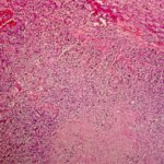
Nasal aspergillosis. Tissue from an 8 year old, neutered male thoroughbred horse with an initial history of sinusitis leading to progressive neurological signs (ataxia, behavioural abnormalities) and prolonged recumbency. Necropsy examination revealed a focus of grey-caseous material within the right nasal chamber that comprised a mat of branching, septae fungal hyphae and mixed inflammatory cells (haematoxylin and eosin stain). Aspergillus spp was cultured from the lesion. There was no gross or histologica

-
Immunofluorescence. Section of renal granuloma from dog J stained with polyclonal antiserum specific for Aspergillus terreus by immunofluorescence.

-
Complement deposition (dog J). Section of myocardial granuloma from dog J stained for canine complement C3 by immunofluorescence. Deposition of C3, but not C4, on fungal hyphae suggests activation of the alternative rather than classical pathway of complement.

-
Lymph node granuloma – Section of lymph node granuloma from a German shepherd dog with disseminated aspergillosis stained for canine IgA by immunofluorescence. The fungal hyphae within the centre of the lesion have surface IgA, and IgA-bearing plasma cells are present within the surrounding inflammatory infiltrate

-
Extensive focus of pyogranulomatous inflammation within the kidney of dog J

-
Aspergillus granuloma within the myocardium of dog J.

-
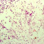
Retinal aspergillosis (dog J) – Section of retina from a German shepherd dog with disseminated aspergillosis. Fungal hyphae and inflammatory cells are found within the vitreous


