Date: 27 January 2014
Copyright:
Fungal Research Trust
Notes:
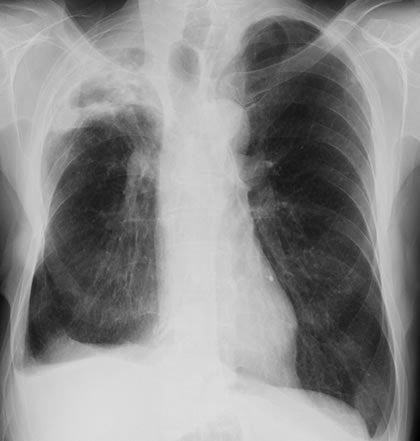
This 73 year old patient with CPA in right upper lobe and COPD who was also a heavy smoker, showed evidence of finger clubbing ( A,B,C). He has been on long term itraconazole, in 1990 he had an oesophagectomy for cancer of the oesophagus. Finger clubbing is an uncommon symptom only seen in advanced or chronic disease. D, chest X ray there are background changes nof COPD with loss of volume in the right hemithorax and a right apical cavititating lesion.
Images library
-
Title
Legend
-
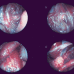
Falcons: The following images were obtained by endoscopy of falcons with aspergillosis.A,B Thoracic airsac (T) with prominent blood vessels and a dead serratospiculum worm (W). The presence of these lung worms makes the airsac look milky. D Normal ovary with developing follicles.

-
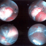
Falcons: The following images were obtained by endoscopy of falcons with aspergillosis.B,D Aspergillus lesions (A) over a swollen liver

-
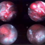
Falcons: The following images were obtained by endoscopy of falcons with aspergillosis.B Cranial, middle, caudal lobes (K1,K2,K3) of the left kidney, all the lobes show slight nephromegaly.C Yellow aspergillus colony (A1), lying adjacent to the lung.D White aspergillus colonies (A2,A3,A4).
-
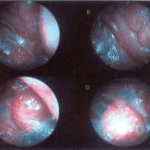
Falcons: The following images were obtained by endoscopy of falcons with aspergillosis.C Cranial pole of left kidney (K) -mildly inflamed.D Ovary ( F) with developing follicles.
-
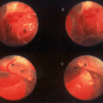
The following images were obtained by endoscopy of falcons with aspergillosis.A and B Lung Worm (S) over liver (Li) (serratospiculum seurati)C and D Aspergilloma (A) and prominent blood vessels on the caudal thoracic air sac (T).
-
The following images were obtained by endoscopy of falcons with aspergillosis. A Lung Worm (serratospiculum sp.) B Lying in betweeen loops of the intestine.C An aspergillus lesion in between the loops of the intestineD Showing cranial pole of left kidney, ovary and oviduct.
