FEV1 spirometry

Date: 7 February 2014

Image B

Copyright:

Dr D Denning, Wythenshawe Hospital, Manchester.(© Fungal Infection Trust)

Notes:

Patient BC
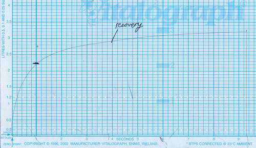
A petite women in her 50’s with severe asthma and fungal sensitization (SAFS) had been unable to tolerate either itraconazole or voriconazole for any length of time, and was severely disabled with her symptoms. One treatment option which is occasionally helpful is to give nebulised amphotericin B (link to video of Helen). She was given 10mg of amphotericin B deoxycholate in water through a Pari LC nebulizer, supervised by a senior physiotherapist. Shortly after starting this, she felt much more breathless and the nebulizer was stopped. Salbutamol rescue was administered. After about 40 minutes she recovered.
 
The spirometer readings show a starting FEV1 of 2.35 L/sec. This fell to 1.05 L/sec, a dramatic fall with amphotericin B nebulisation. (Image A) She recovered with salbutamol to 2.25 L/sec. (Image B)


Images library

Showing 10 posts of 2574 posts found.
  • Title

    Legend

  • slide9pat_AB

  • slide10pat_AB

  • slide11pat_AB

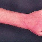
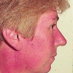
  • Facial erythema: Voriconazole rash in ABPA patient resistant to corticosteroids, treated with voriconazole 200mg BID. Serum voriconazole levels were very low and the dose was raised to 250mg BID. Within 3 weeks patient had developed remarkable facial erythema. His trough voriconazole concentration at this time was 370ng/ml. When voriconazole was stopped because of the facial erythema and lack of impact on his ABPA his facial erythema resolved over 4 weeks.

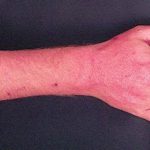
    Forearm erythema related to voriconazole. As with facial erythema patient developed remarkable forearm erythema with lesions similar to porphyria cutanea tarda all of which resolved with stopping voriconazole.

    Facial erythema related to voriconazole, Forearm erythema related to voriconazole 1, Forearm erythema related to voriconazole 2

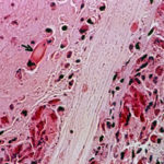
  • Eosinophilic mucin containing numerous eosinophils and Charcot-Leyden crystals (arrow). Stain PAS x400. Patient with allergic fungal sinusitis
    SJTflavus1

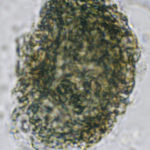
  • A whole fungal ball removed from the sinus by endoscopic surgery. No staining x 10
    wholefungusball

  • Crushed fungal material removed from sinus by endoscope. No staining x40

    crushedfungusball

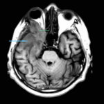
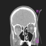
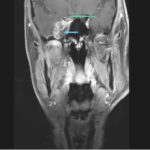
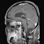
  • This 68 year man with a history of hypertension and ischemic heart disease presented with nasal obstruction, localised swelling and pain in his right cheek for about two months. CT scan showed a soft tissue mass filling the right maxillary sinus adjacent to the floor of the orbit. Maxillotomy with mass removal was performed and culture grew A. fumigatus. Histology was not performed and the patient received no antifungal therapy. 5 months later localised relapse with progression along the medial wall of the orbit was seen on CT scan.
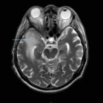
    Image A. MRI (T2-weighted, transversal view).Note oedema of the right temporal lobe., Image B MRI (T1-weighted, transversal view). The blue pointer shows progression of inflammatory tissue into the brain. The green pointer shows involvement of the lateral group of external ocular muscles., Image C CT scan image (bone window, coronal view) demonstrating destruction of the inferior wall of the right orbit., Image D. MRI (T1-weighted, contrast-enhanced, transversal view). The pointers show abnormal enhancement in the right orbit (green), in the right temporal lobe (blue) and of the dura (yellow). , Image E MRI (T1-weighted, contrast-enhanced, coronal view). The pointers show pathological tissue in the right cavernous sinus (blue) and pathological enhancement of the right optical nerve (green)., Image F MRI (T1-weighted, contrast-enhanced, sagittal view). The pointers show pathological tissue in the right orbit (blue) with protrusion into the right optical canal (green).

  • Yamik catheter for rinsing nasal and paranasal cavities. Image D

    xray2

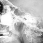
  • Yamik catheter for rinsing nasal and paranasal cavities. Image C
    xray1