Date: 26 November 2013
Fig3 Pulmonary artery
Copyright: n/a
Notes:
Fig 1. Trachea and bronchi A 50+ year old woman received a double lung transplant for emphysema. She did well initially, but then Aspergillus fumigatus was grown from her airways, in association with mucous and a pseudomembrane covering parts of her anastomosis and airways. 2 months after her transplant she was undergoing bronchoscopy, and started to bleed. This rapidly became torrential and she suffered a cardiac arrest and died.
She underwent autopsy at which it was found that the larynx, trachea and major bronchi all contained blood (Fig 1).
The bronchial anastomoses were intact, but brown fluffy material was found overlying the stitches on both sides. On the right side plaques of similar material were seen distal to the anastomoses, overlying an ulcer and an obstructing the smaller bronchi.
On the left side an ulcer 1.5cm in diameter (with blood in it) was seen in the main bronchus distal to the anastomosis on the anterior wall (Fig 2). The sutures of the anastomosis are intact. The centre of the ulcer had ulcerated through into the left main pulmonary artery (Fig 3). The pulmonary artery shows necrosis and discolouration of the intimal surface over an area of 1.5-1.0cm.
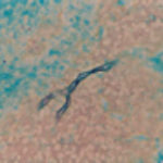
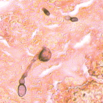
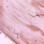
Histopathology examination showed fungal hyphae perforating the bronchial wall and arterial wall around and in the ulcer. The ulcer on the right side showed hyphae perforating the wall and bronchial cartilage.
Images library
-
Title
Legend
-
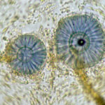
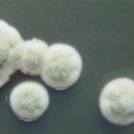
Scanning electron micrograph of Aspergillus ochraceopetaliformis conidial heads
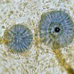
-
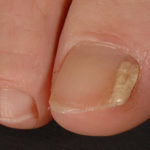
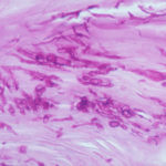
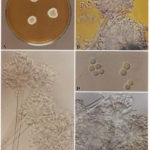
Image D & E. A case of onychomycosis associated with Aspergillus ochraceopetaliformis as described in Nail infection by Aspergillus ochraceopetaliformis. Med Mycol. 2009 Mar 9:1-5, 2009, Brasch J, Varga J, Jensen JM, Egberts F & Tintelnot K
 ,
,  ,
,  ,
,  ,
, 
-
Further details
Image 5. Oral itraconazole pulse therapy was given to the patient (200 mg twice daily for 1 week, with 3 weeks off between successive pulses, for four pulses) and treatment was successful.
 ,
,  ,
,  ,
,  ,
, 
-
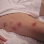
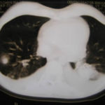
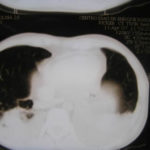
This patient was 28 yr old with adult lymphocytic leukaemia. She received induction chemotherapy and this infection developed 2 days after recovering from neutropenia.
 ,
,  ,
,  ,
,  ,
,  ,
,  ,
,  ,
,  ,
,  ,
, 
-
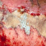
Close-up image of the lesion on the left thigh showing a mat of hyphae over the wound.
-
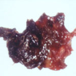
Eosinophilic mucin with A. flavus in the nasal cavity. Irregular crust of 2.5 cm from a patient diagnosed as allergic fungal sinusitis. Patient with allergic fungal sinusitis
-
GMS stain of eosinophilic mucin reveals a darkly stained dichotomously branched A. flavus hyphae within cellular background. Patient with allergic fungal sinusitis
