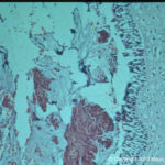
Date: 26 November 2013
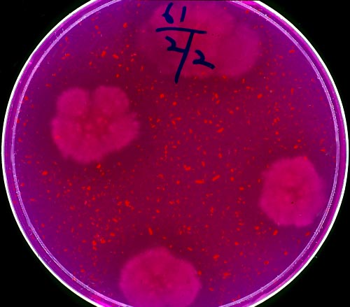
Four colonies of Aspergillus on an agar plate containing rose bengal (to limit colony spending) and elastin fibres (light pink dots). Underneath and surrounding the colonies, the elastin fibres have gone, indicating enzymatic degradation
Copyright:
© Denning DW, Elliott J, Keaney M. Temperature-dependent expression of elastase in Aspergillus species. J Med Vet Mycol, 1993;31:455-458.
Notes:
The medium was described by: Kothary MH, Chase T, Macmillan JD. Correlation of elastase production by some strains of Aspergillus fumigatus with ability to cause pulmonary invasive aspergillosis in mice. Infect Immun 1984; 43:320-3235.
Images library
-
Title
Legend
-
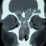
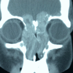
4 Total obstruction of the sinuses due to inflamed mucosa. (Patient 04)
-
1 Axial computed tomography (CT) scans of the frontal sinus.
A: due to the long lasting pressure of mucus, the bone of the anterior wall of frontal sinus is thinned out and elevated anteriorly, forming a bulge. B: same situation as depicted in fig A: the posterior bony wall of frontal sinus is thinned out and extremely elevated posteriorly towards the frontal lobe of the brain. As depicted on the scan, a thin bony layer covering the dura could be recognized intraoperatively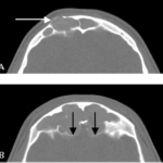
-
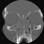
2 Same patient as 1 and 3, frontal CT
-
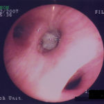
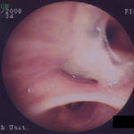
D. 6 months later, tenacious yellow secretions in L basal bronchial division
-
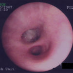
C. After suction the material was seen to extend distally – obstructing the right basal stem bronchus
-
B. After suction the material was seen to extend distally – obstructing the right basal stem bronchus
