Date: 26 November 2013
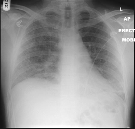
22/09/08 This chest radiograph shows bilateral hazy diffuse airspace disease predominating in the lower lungs with subtle nodularity in upper zones.
Copyright: n/a
Notes:
A 33 year old known Chronic Granulomatous Disorder (CGD) male presented to A&E in respiratory distress and admitted with severe bibasal pneumonia. He had been laying mulch in his garden. He had not been taking any prophylactic antifungal agents. Oxygen therapy was commenced in conjunction with IV bacterial and fungal treatment with Amphotericin B (Fungizone ®). Further consultation and an adverse reaction to the administration of Fungizone ® led to a switch to IV Voriconazole 300mg BD. The patient tested positive for aspergillus antibodies in serum. The patient declined a bronchoscopy, responded well to IV voriconazole and was discharged home 2 weeks post admission on maintenance voriconazole.
Images library
-
Title
Legend
-
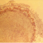
The periphery of the fungus ball is deeply eosinophilic because of the deposition of Splendore-Hoeppli material.
-
Single fungal ball, moving. Radiographic appearance of a fungus ball, showing movement as the patient’s position changes.
-
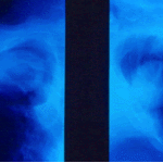
Oxalate crystals in the cavity wall surrounding an Aspergillus niger fungus ball (H&E, dark field, x 25).
-
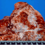
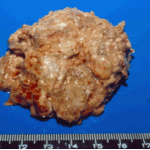
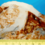
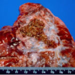
Aspergilloma patient. Gross pathology appearance of a fungus ball.
-
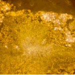
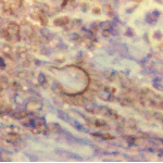
Conidiophores of Aspergillus fumigatus in the mass of the fungal ball surrounded by mycelia (H&E, x 400).
-
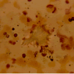
Aspergillus niger fungal ball. Calcium oxalate crystals in Aspergillus niger fungal ball. Also shown are darkly pigmented, rough-walled conidia associated with Aspergillus niger infection.
-
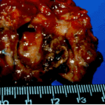
Aspergillus niger fungus ball within an old tuberculous cavern. This patient had diabetes, a disease commonly associated with A. niger infection.

 ,
, 
 ,
, 