Date: 26 November 2013
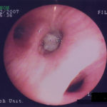
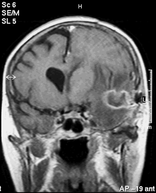
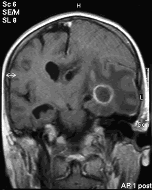
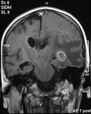
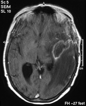
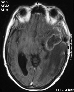
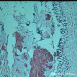
T1 weighted, gadolinium enhanced magnetic resonance brain scan. This 43 year-old alcoholic woman underwent laparoscopic cholecystectomy in January 2001. Ten days after surgery, she became confused, dysphasic and eventually had tonic-clonic seizures. A CT scan showed non-communicating hydrocephalus with ventriculitis. She underwent many complicated neurosurgical interventions, and received long term broad-spectrum antimicrobials and dexamethasone. One month later, she had generalized seizures, and a large abscess was observed on scan (see images). A heavy growth of A. fumigatus was retrieved from the abscess, and amphotericin B and 5-flucytosine were started. Antifungal therapy was changed voriconazole due to intolerance to amphotericin B and worsening disorientation. Voriconazole dosing (which varied from 300mg to 100mg twice daily) was guided by plasma concentrations as enzyme induction with rifampicin and carbamazepine, and reduction in clearance with alcoholic liver disease complicated her voriconazole dosing. Steroids were gradually reduced. She had a good recovery and completed 9 months of voriconazole.
Despite air filtration in the operating rooms, she apparently acquired an intra-operative infection, probably accelerated in presentation by concurrent dexamethasone. Rapid diagnosis and optimization of voriconazole dosing lead to a good outcome.
Copyright: n/a
Notes: n/a
Images library
-
Title
Legend
-
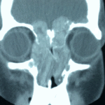
4 Total obstruction of the sinuses due to inflamed mucosa. (Patient 04)
-
1 Axial computed tomography (CT) scans of the frontal sinus.
A: due to the long lasting pressure of mucus, the bone of the anterior wall of frontal sinus is thinned out and elevated anteriorly, forming a bulge. B: same situation as depicted in fig A: the posterior bony wall of frontal sinus is thinned out and extremely elevated posteriorly towards the frontal lobe of the brain. As depicted on the scan, a thin bony layer covering the dura could be recognized intraoperatively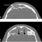
-
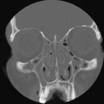
2 Same patient as 1 and 3, frontal CT
-
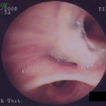
D. 6 months later, tenacious yellow secretions in L basal bronchial division
-
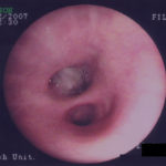
C. After suction the material was seen to extend distally – obstructing the right basal stem bronchus
-
B. After suction the material was seen to extend distally – obstructing the right basal stem bronchus
