Date: 26 November 2013
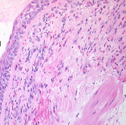
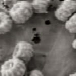
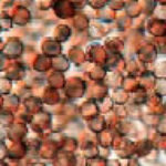
Bronchial mucosa under H & E stain showing numerous eosinophils deep to the mucosa, and mucus in the lumen of the bronchiole.
Copyright:
Fungal Infection Trust
Notes:
Mucoid impaction due to ABPA- Pt DL.A 57 year old woman presented with breathlessness. She had a history of mild asthma for which she occasionally took salbutamol inhaler puffs. The patient underwent a pneumonectomy because of the severity of her disease process, and uncertainty about the diagnosis, prior to serology results being obtained.Serology showed an IgE of 2600, with a strongly positive Aspergillus RAST test and weakly positive Aspergillus precipitins. Material removed at bronchoscopy showed eosinophilia. These features confirm a diagnosis of allergic bronchopulmonary aspergillosis (ABPA).
Images library
-
Title
Legend
-
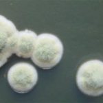
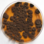
Pigmentation of Aspergillus versicolor colonies ranged from pale green to greenish-beige, pink-green, dark green and brown. Reverse is usually reddish. The growth rate is usually slow. Cultured on Sabouraud dextrose agar with chloramphenicol.
-
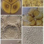
A Colonies on MEA after one week; B, C conidial heads with tip of conidiophire, x920; D conidial head, x 2330; E conidial heads x920
![aspvers[2] aspvers2](https://www.aspergillus.org.uk/wp-content/uploads/2017/10/aspvers2-150x150.jpg)
-
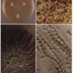
A Colonies on MEA + 20% sucrose after one week; B detail of colony showing columnar conidial heads x 44 ; C conidial heads x 920; D conidia x2330
-
Cultures are grown on malt extract agar for 5-7 days at 30°C.
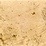
Light microscopy-1000x stained with lacto-phenol and cotton blue.
-
A Colonies on MEA +20% sucrose after one week; B ascomata x 40; C conidiophores x 920; D ascospores x2330; E ascoma x 230; F portion of ascoma with asci and ascospores, x 920.
