Date: 26 November 2013
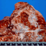
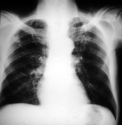
Bilateral upper-lobe cavities in AIDS, pt PC
Copyright: n/a
Notes:
This patient, thought initially to have pulmonary aspergillosis in AIDS, has bilateral upper-lobe cavities, more marked on the left. He presented with fever, nonproductive cough and dyspnoea. Bronchoscopy yielded Aspergillus fumigatus. He refused therapy and died with progressive disease. He is reported as patient 8 in Denning DW, Follansbee S, Scolaro M, Norris S, Edelstein D, Stevens DA. Pulmonary aspergillosis in AIDS. N Engl J Med 1991; 324: 654-662.
Images library
-
Title
Legend
-
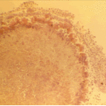
The periphery of the fungus ball is deeply eosinophilic because of the deposition of Splendore-Hoeppli material.
-
Single fungal ball, moving. Radiographic appearance of a fungus ball, showing movement as the patient’s position changes.
-
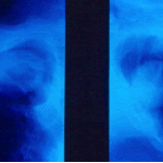
Oxalate crystals in the cavity wall surrounding an Aspergillus niger fungus ball (H&E, dark field, x 25).
-
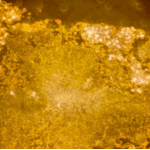
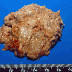
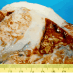
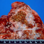
Aspergilloma patient. Gross pathology appearance of a fungus ball.
-
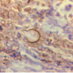
Conidiophores of Aspergillus fumigatus in the mass of the fungal ball surrounded by mycelia (H&E, x 400).
-
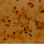
Aspergillus niger fungal ball. Calcium oxalate crystals in Aspergillus niger fungal ball. Also shown are darkly pigmented, rough-walled conidia associated with Aspergillus niger infection.
-
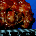
Aspergillus niger fungus ball within an old tuberculous cavern. This patient had diabetes, a disease commonly associated with A. niger infection.

 ,
, 
 ,
, 