Date:
Copyright:
TAIJIRO TOMIKAWA, KAZUO SHIN-YA, HARUO SETO, NORIYUKI OKUSA, TAKAYUKI KAJIURA, YOICHI HAYAKAWA, Structure of Aspochalasin H, a New Member of the Aspochalasin Family, The Journal of Antibiotics, 2002, Volume 55, Issue 7, Pages 666-668, Released on J-STAGE January 27, 2009, Online ISSN 1881-1469, Print ISSN 0021-8820, https://doi.org/10.7164/antibiotics.55.666, https://www.jstage.jst.go.jp/article/antibiotics1968/55/7/55_7_666/_article/-char/en
Notes:
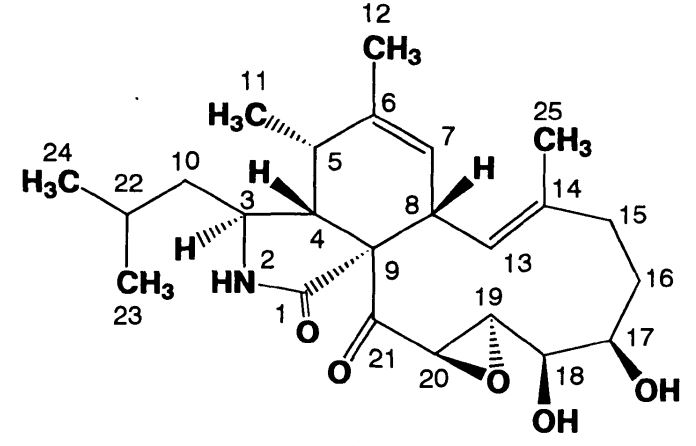
Structure of Aspochalasin H
Images library
-
Title
Legend
-
Single fungal ball, moving. Radiographic appearance of a fungus ball, showing movement as the patient’s position changes.
-
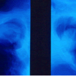
Oxalate crystals in the cavity wall surrounding an Aspergillus niger fungus ball (H&E, dark field, x 25).
-
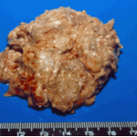
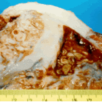
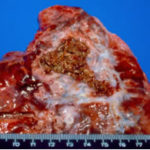
Aspergilloma patient. Gross pathology appearance of a fungus ball.
-
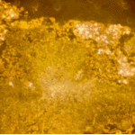
Conidiophores of Aspergillus fumigatus in the mass of the fungal ball surrounded by mycelia (H&E, x 400).
-
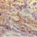
Aspergillus niger fungal ball. Calcium oxalate crystals in Aspergillus niger fungal ball. Also shown are darkly pigmented, rough-walled conidia associated with Aspergillus niger infection.
-
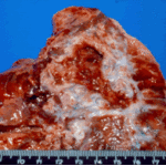
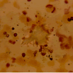
Aspergillus niger fungus ball within an old tuberculous cavern. This patient had diabetes, a disease commonly associated with A. niger infection.
-
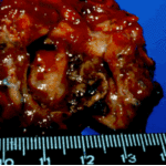
Conidial head and brown conidia in a section of a fungus ball caused by Aspergillus niger (H&E, x 400).


 ,
, 
 ,
, 